

Fluo-3, AM Ester, 1Mm In Anhydrous Dmso

Référence 50015
Conditionnement : 1ML
Marque : Biotium
Fluo-3, AM Ester, 1 mM in Anhydrous DMSO
Membrane-permeant AM ester form of Fluo-3 that can be loaded into cells via incubation. Because of the relatively low water solubility of the AM ester, the mild detergent Pluronic F-127 (catalog no. 59004) is often used as a dispersing agent to facilitate loading.
Product Description
Membrane-permeant AM ester form of Fluo-3 that can be loaded into cells via incubation. Because of the relatively low water solubility of the AM ester, the mild detergent Pluronic® F-127 (cat# 59004) is often used as a dispersing agent to facilitate loading. Fluo-3 AM ester itself does not bind Ca2+, but it is readily hydrolyzed to Fluo-3 by endogenous esterases once the dye is inside the cells. An important application of Fluo-3 AM ester is its use in high throughput drug screening. For information on Fluo-3, see cat# 50010.
- Orange red liquid
- Store -20°C and protect from light
- C51H50Cl2N2O23
- MW: 1129.9
- [121714-22-5]
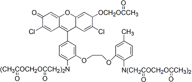
BAPTA-based ion indicators like Fluo-3 have been shown to be fixable in situ by EDC/EDAC (cat# 59002). The fixation of indicator dyes is useful for downstream immunofluorescence and IHC studies (Cell Calcium 1997, 21(3), 175).
As the indicator does not covalently bind to cellular components, it may be actively effluxed from the cell by organic anion transporters. The rate of efflux increases with temperature, and may vary between cell types, resulting in variable retention times of a few minutes to hours. Experiments using indicators in cells usually are performed within one or two hours of loading, but it may be possible to re-load cells with indicator if needed. The organic anion transporter inhibitor Probenecid (#50027) can be used to slow the rate of indicator efflux from cells.
Technology

Product Attributes
CAS number
Size
Cell permeability
Indicator type
Excitation/Emission
Documents, Protocols, SDS and COA

Protocols
SDS
COA
Request the Certificate of AnalysisReferences
- Methods Cell Biol, 99, 113, (2021), DOI: 10.1016/B978-0-12-374841-6.00005-0