| Catalog Number | Cell Type | IPSC Line | Sex | Reporter | Edit | 1M Size Vial | 2M Size Vial | 5M Size Vial | hf:categories | hf:att:pa_ipsc-line | hf:att:pa_sex | hf:att:pa_reporter | hf:att:pa_edit |
|---|---|---|---|---|---|---|---|---|---|---|---|---|---|
| BX-0900-30 | Microglia | WC-30 | M | Add to cart | Add to cart | Add to cart | microglia | wc-30 | m | ||||
| BX-0900-32 | Microglia | WC-32 | F | Add to cart | Add to cart | Add to cart | microglia | wc-32 | f | ||||
| BX-0914-32 | Microglia | WC-32 | F | APOE4/E4 | Add to cart | Add to cart | Add to cart | microglia disease-cells | wc-32 | f | apoe4-e4 | ||
| BX-0920-32 | Microglia | WC-32 | F | TREM2 KO | Add to cart | Add to cart | Add to cart | microglia disease-cells | wc-32 | f | trem2-ko | ||
| BX-0921-32 | Microglia | WC-32 | F | TREM2 R47H | Add to cart | Add to cart | Add to cart | microglia disease-cells | wc-32 | f | trem2-r47h | ||
| BX-0900-33 | Microglia | BX-33 | F | Add to cart | Add to cart | Add to cart | microglia | bx-33 | f | ||||
| BX-0901-30 | Microglia | WC-30 | M | eGFP | Add to cart | Add to cart | Add to cart | microglia reporter-cells | wc-30 | m | egfp | ||
| BX-0922-32 | Microglia | WC-32 | F | TREM2 R62H | Add to cart | Add to cart | Add to cart | microglia | wc-32 | f | trem2-r62h | ||
| BX-0923-32 | Microglia | WC-32 | F | TREM2 D87N | Add to cart | Add to cart | Add to cart | microglia | wc-32 | f | trem2-d87n |
Overview
| Product ID | BX-0900 |
|---|---|
| Description | BrainXell Microglia are cryopreserved human iPSC-derived microglia available from male and female lines, including WT, reporter, and disease-associated variants such as APOE4/E4, TREM2 KO, TREM2 R47H, TREM2 R62H, and TREM2 D87N. These cells are highly effective for neuroinflammation studies, as they respond to external pro-inflammatory factors by secreting well-known cytokines. Furthermore, they are capable of phagocytosing β-amyloid, making them a versatile tool for disease modeling and integration into neuron-glia co-culture workflows. |
Maturation
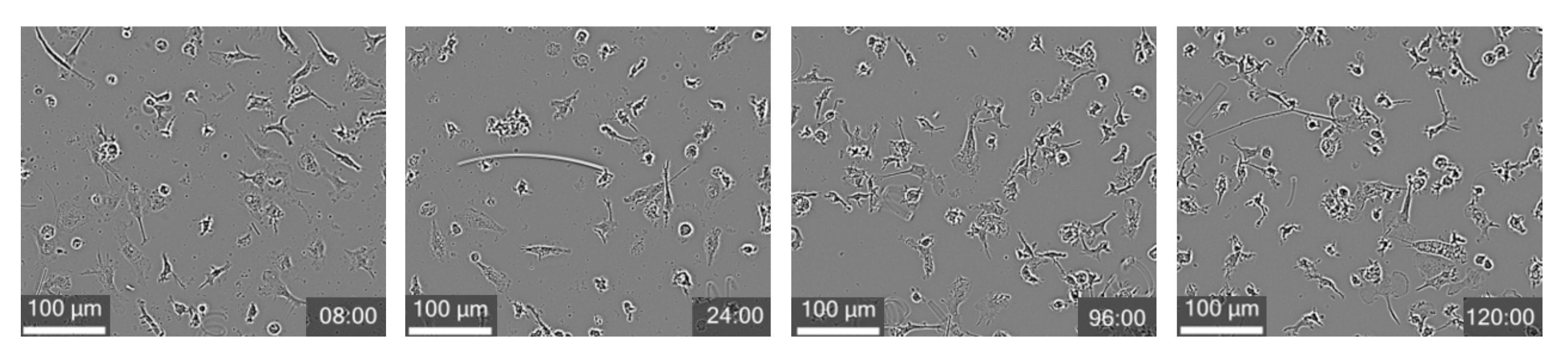
- BrainXell Microglia develops rapidly in vitro, achieving established phenotypic characteristics such as transitioning from rounded or amoeboid shapes into increasingly ramified structures.
Reproducibility
- BrainXell Microglia are generated using standardized differentiation and QC protocols, resulting in consistent marker expression and functional performance across experiments and culture formats.
Their compatibility with standardized plate formats and controlled assay conditions further enables reproducible, scalable workflows for screening and downstream applications.
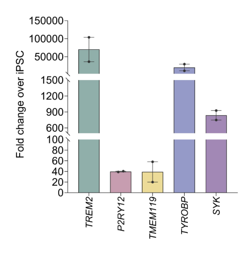
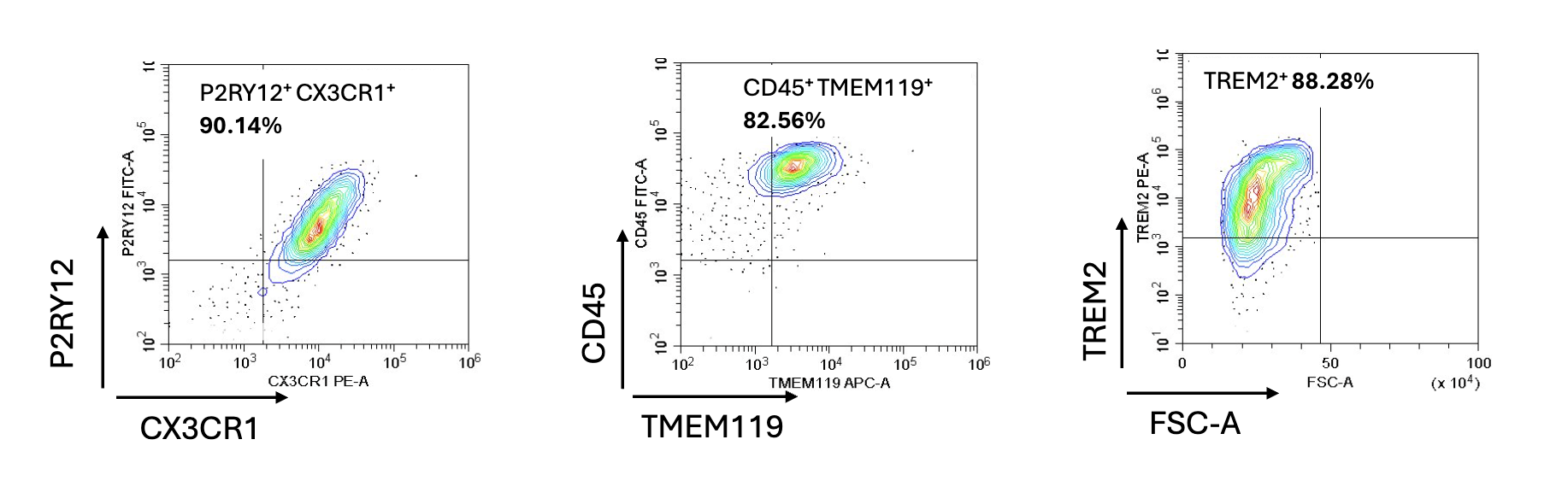
Expression
BrainXell Microglia express canonical microglial markers including CD45, CD11b, CX3CR1, P2RY12, TMEM119, TREM2, and IBA1. Flow cytometry data show strong expression of P2RY12/CX3CR1-positive cells at ~90%, CD45/TMEM119-positive cells at ~83%, and TREM2-positive cells at ~88%.
Functionality
BrainXell Microglia can be stimulated with known pro-inflammatory agents to secrete cytokines. BrainXell microglia also demonstrate β-amyloid phagocytosis across WT and disease-relevant genotypes.
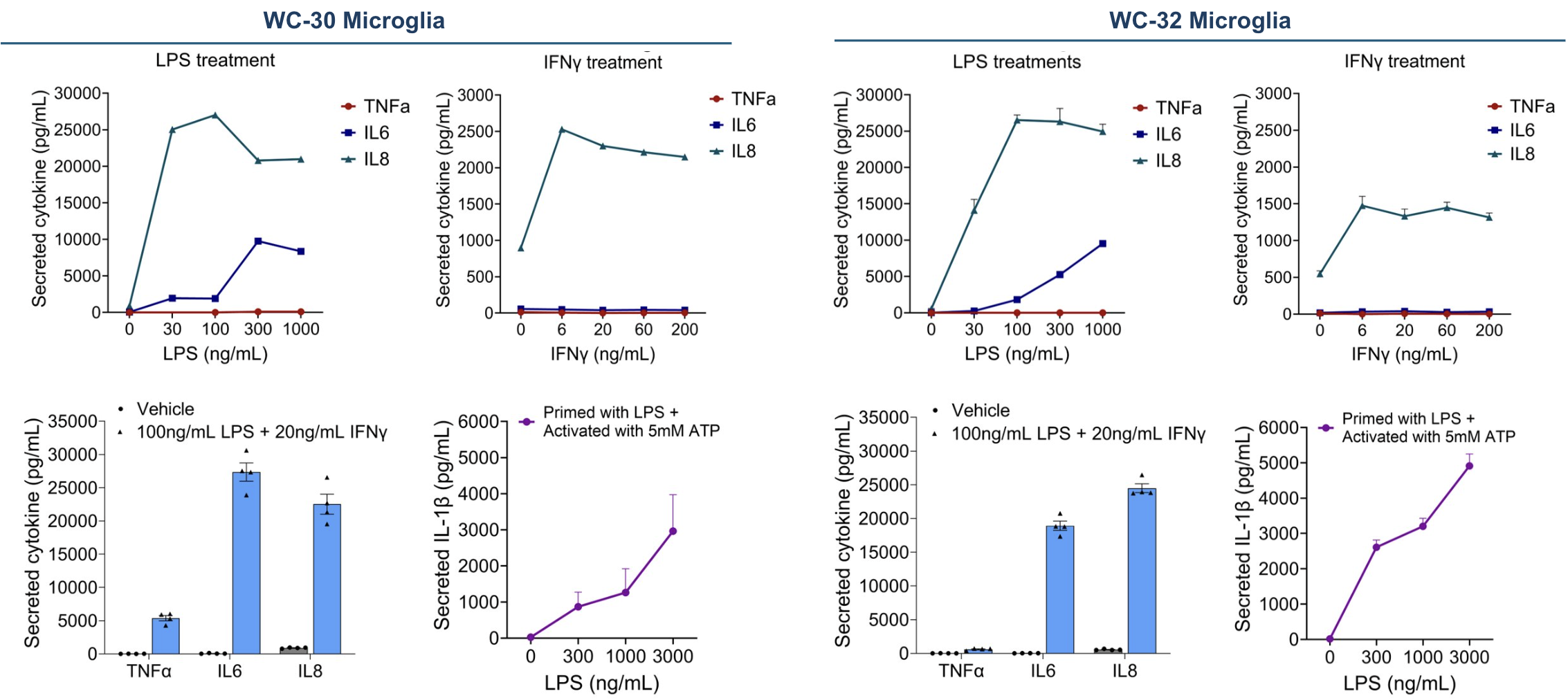
Cytokine secretion
BrainXell WC-30 (male iPSC line) and WC-32 (female iPSC line) microglia were cultured for 7 days post-thaw. IL-1β secretion was measured following priming with the indicated concentrations of LPS for 3 hours, followed by activation with 5 mM ATP for 30 minutes. Secretion of TNFα, IL-6, and IL-8 cytokines was measured after 48 hours of treatment with the indicated conditions (at day 9 post-thaw).
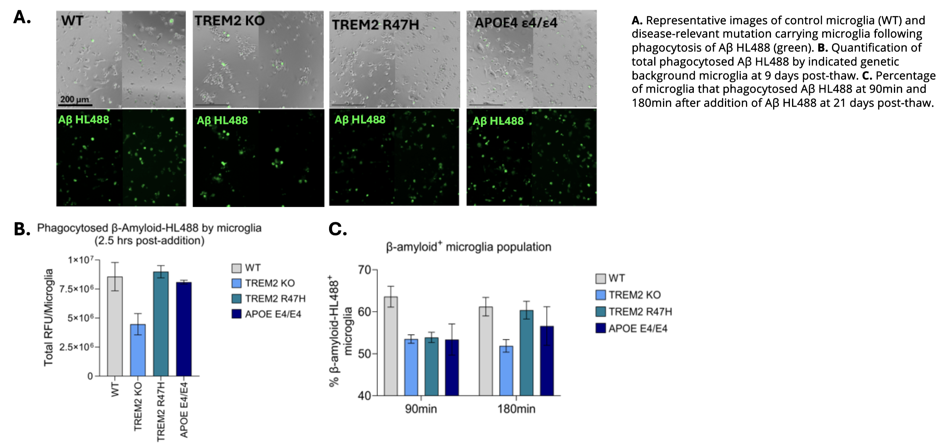
Phagocytosis
BrainXell Microglia demonstrate quantifiable β-amyloid phagocytosis across WT and disease-relevant genotypes, including TREM2 KO, TREM2 R47H, and APOE E4/E4 variants. Time-course analyses show active uptake of fluorescent Aβ over multiple hours, supporting applications in neurodegeneration research, microglial activation studies, and functional screening workflows focused on Alzheimer’s disease.
Applications
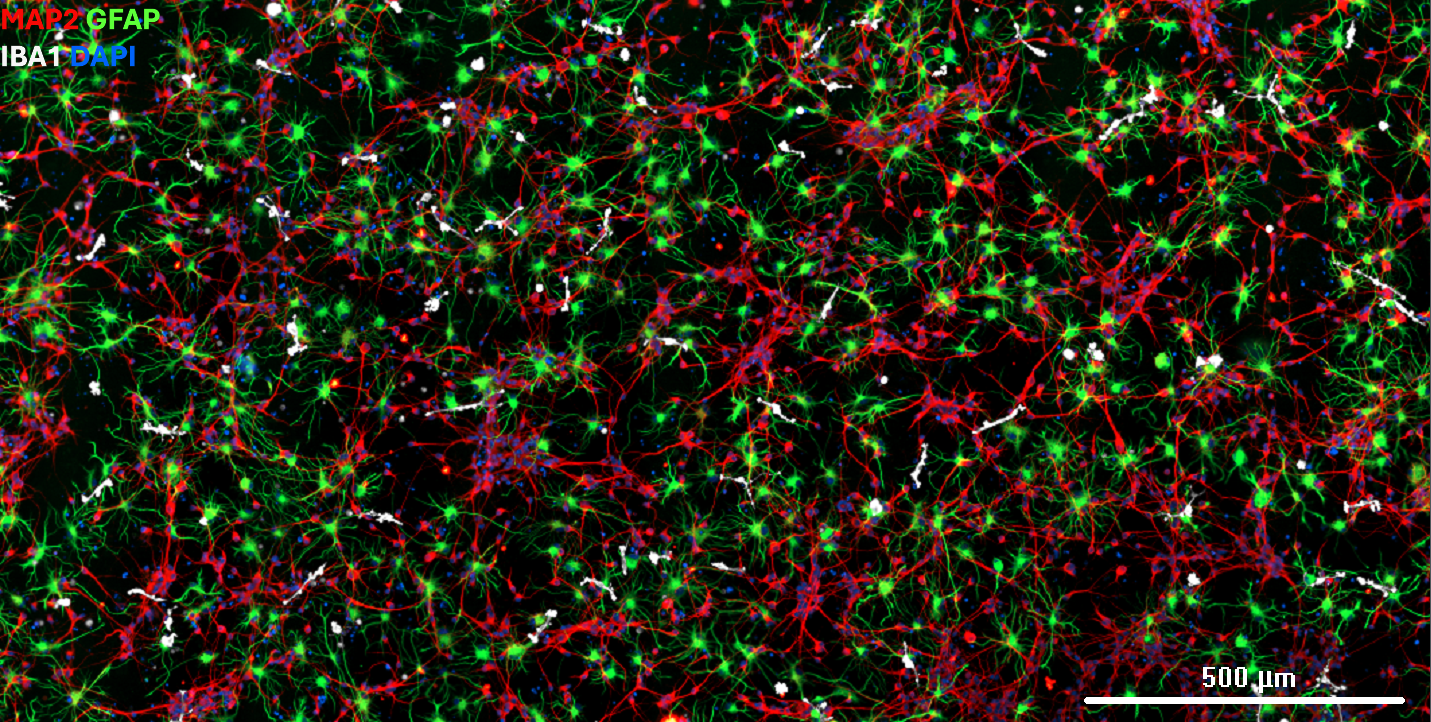
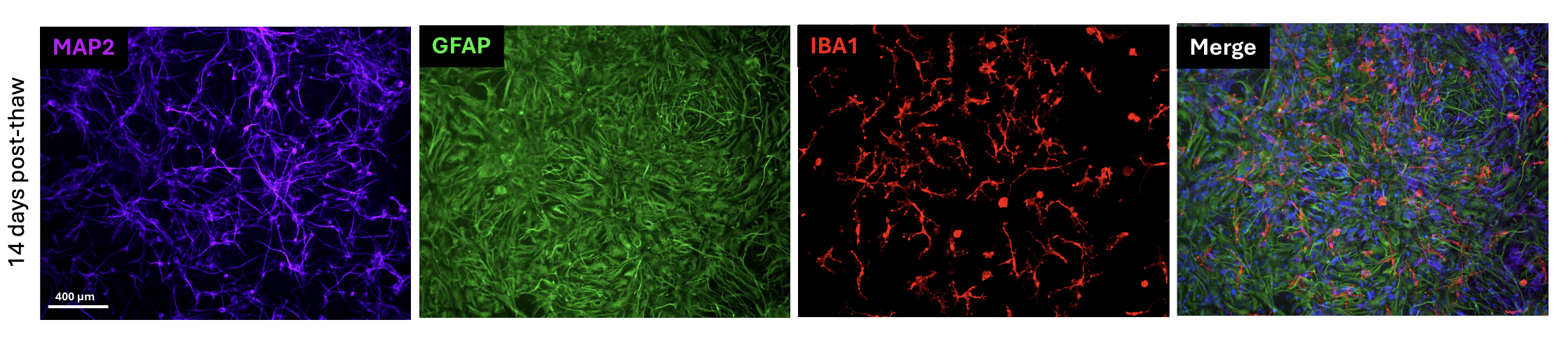
BrainXell iPSC-derived cortical glutamatergic neurons, cortical astrocytes, and microglia can be established in a tri-culture system that more closely recapitulates the physiological cellular environment and neural network. In this model, immunostaining at 14 days post-thaw demonstrates that microglia adopt a mature, ramified morphology closely resembling
that observed in vivo.
Efficient gene delivery to BrainXell iPSC-derived microglia can be achieved using lipid nanoparticle (LNP)-based transfection methods. Please contact us for an application note describing this workflow in detail.
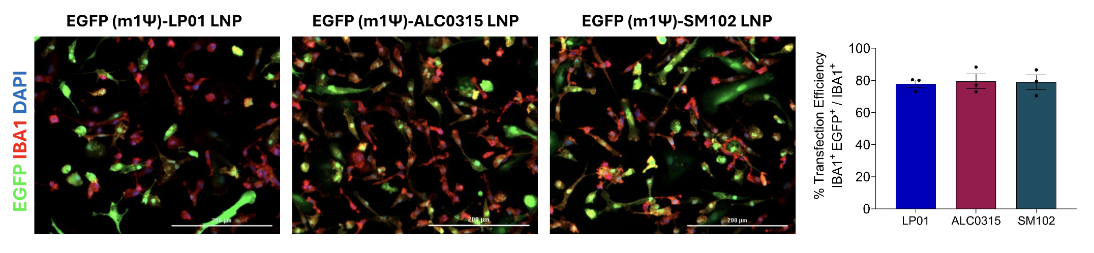
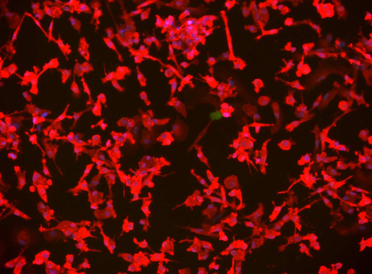
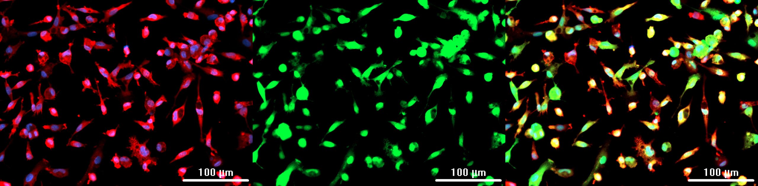
BrainXell Microglia were transfected with EGFP (m1Ψ) mRNA containing different types of LNPs at 4 days post-seeding. Cultures were maintained for 7 days after transfection (day 11 post- thaw) and fixed. Immunostaining was performed for IBA1 (microglia marker) and DAPI (nuclear stain). Scale bar: 200 µm. Quantification of transfection efficiency in microglia was determined by dividing IBA1 and EGFP double positive cell count by the total IBA1 positive cell count in 3 independently transfected wells.
Protocols
BrainXell recommends a dedicated Microglia Culture Protocol for mono-culture maintenance, with additional protocols available for Neuron-Microglia co-culture and Neuron-Astrocyte-Microglia tri-culture workflows. Co-culture protocol recommendations may vary depending on application, and BrainXell application scientists can help guide experimental design.
BrainXell Microglia Monoculture Protocol v10.2
BrainXell Co-culture of Neuron_Microglia Protocol v11.0
BrainXell Tri-Culture of Neuron_Astrocyte_Microglia Protocol v11.0