


Anti-Thomsen-Friedenreich Antigen / CD176 (Pan Carcinoma Marker) Monoclonal Antibody(Clone: A63-C/A9)
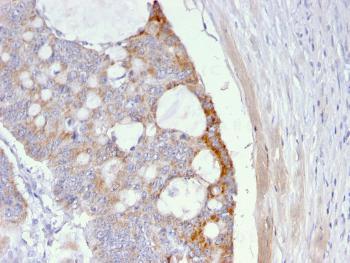
Fig. 1: Formalin-fixed, paraffin-embedded human Colon Carcinoma stained with T-F Antigen Mouse Monoclonal Antibody (A63-C/A9).
| Amount : | 100 µg |
| Isotype : | Mouse IgM, kappa |
| Content : | 200 µg/ml of Ab Purified from Bioreactor Concentrate. Prepared in 10mM PBS with 0.05% BSA & 0.05% azide. Also available WITHOUT BSA & azide at 1.0mg/ml. |
| Storage condition : | Antibody with azide - store at 2 to 8°C. Antibody without azide - store at -20 to -80°C. Antibody is stable for 24 months. Non-hazardous. |
| Alternative Name : | T-F Antigen; TF Antigen; Asialoglycophorin; pan-carcinoma marker; CD176 |
| Immunogen Information : | Neuraminidase-treated human red blood cells |
Recognizes a disaccharide epitope, Gal 1-3GalNAc, of Thomsen-Friedenreich (TF) antigen. It is specific for both anomeric forms of the disaccharide (TF and TF , including related structures on the glycolipid) and shows no cross-reactivity with sialylated glycophorin. The Thomsen-Friedenreich antigen acts as an oncofetal antigen, with low expression in normal adult tissues but increasing to fetal levels of expression in hyperplasia or malignancy. It is considered as a pan-carcinoma marker. This MAb is capable to agglutinate desialylated red blood cells. During metastasis, the ability of malignant cells to form multicellular aggregates via homotypic or heterotypic aggregation and their adhesion to the endothelium are critical. The tumor-associated carbohydrate Thomsen-Friedenreich antigen (Gal-GalNAc) is involved in tumor cell adhesion and tissue invasion. It also causes an immune response, and overexpression of the antigen causes cancer cells to be more sensitive to natural killer cell lysis. The Thomsen-Friedenreich antigen is suppressed in normal healthy cells and represents one of the few chemically well-defined antigens associated with tumor malignancy. The presence of the Thomsen-Friedenreich antigen on the surface of cancer cells may result from a divergence from the normal pathway for O-linked glycosylation in these cells, most likely caused by inappropriate localization of the enzymes involved in synthesis of the disaccharide.
Immunofluorescence (0.5-1µg/ml); Immunohistochemistry (Frozen) (0.5-1.0µg/ml for 30 minutes at RT); Immunohistochemistry (Formalin-fixed) (1-2µg/ml for 30 minutes at 37'C)(Staining of formalin-fixed tissues requires boiling tissue sections in 10mM citrate buffer, pH 6.0, for 10-20 min followed by cooling at RT for 20 minutes)Optimal dilution for a specific application should be determined.
For Research Use Only. Not for use in diagnostic/therapeutics procedures.
| There are currently no product reviews |