

Anti-CYP1A1 Antibody

Katalog-Nummer MBS178240-0,1mg
Size : 0.1mg
Marke : MyBiosource
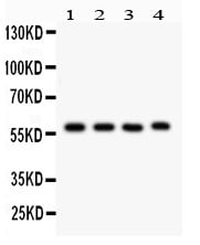
Figure 1. Western blot analysis of CYP1A1 using anti-CYP1A1 antibody (MBS178240).
Electrophoresis was performed on a 5-20% SDS-PAGE gel at 70V (Stacking gel) / 90V (Resolving gel) for 2-3 hours. The sample well of each lane was loaded with 50ug of sample under reducing conditions.
Lane 1: Rat Lung Tissue Lysate
Lane 2: Mouse Lung Tissue Lysate
Lane 3: Human Placenta Tissue Lysate
Lane 4: JURKAT Whole Cell Lysate
After Electrophoresis, proteins were transferred to a Nitrocellulose membrane at 150mA for 50-90 minutes. Blocked the membrane with 5% Non-fat Milk/ TBS for 1.5 hour at RT. The membrane was incubated with rabbit anti-CYP1A1 antigen affinity purified polyclonal antibody at 0.5ug/mL overnight at 4 degree C, then washed with TBS-0.1%Tween 3 times with 5 minutes each and probed with a goat anti-rabbit IgG-HRP secondary antibody at a dilution of 1:10000 for 1.5 hour at RT. The signal is developed using an Enhanced Chemiluminescent detection (ECL) kit with Tanon 5200 system. A specific band was detected for CYP1A1 at approximately 58KD. The expected band size for CYP1A1 is at 58KD.
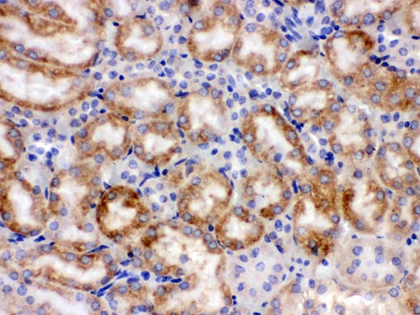
Figure 2. IHC analysis of CYP1A1 using anti-CYP1A1 antibody (MBS178240).
CYP1A1 was detected in paraffin-embedded section of Mouse Kidney Tissue. Heat mediated antigen retrieval was performed in citrate buffer (pH6, epitope retrieval solution) for 20 mins. The tissue section was blocked with 10% goat serum. The tissue section was then incubated with 1ug/ml rabbit anti-CYP1A1 Antibody (MBS178240) overnight at 4 degree C. Biotinylated goat anti-rabbit IgG was used as secondary antibody and incubated for 30 minutes at 37 degree C. The tissue section was developed using Strepavidin-Biotin-Complex (SABC) with DAB as the chromogen.
CYP1A1 was detected in paraffin-embedded section of Mouse Kidney Tissue. Heat mediated antigen retrieval was performed in citrate buffer (pH6, epitope retrieval solution) for 20 mins. The tissue section was blocked with 10% goat serum. The tissue section was then incubated with 1ug/ml rabbit anti-CYP1A1 Antibody (MBS178240) overnight at 4 degree C. Biotinylated goat anti-rabbit IgG was used as secondary antibody and incubated for 30 minutes at 37 degree C. The tissue section was developed using Strepavidin-Biotin-Complex (SABC) with DAB as the chromogen.
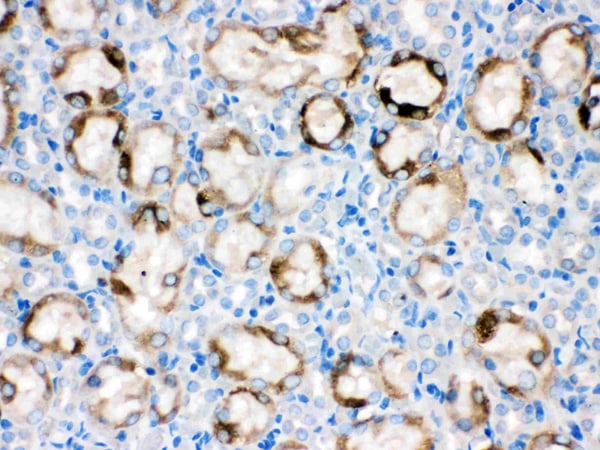
Figure 3. IHC analysis of CYP1A1 using anti-CYP1A1 antibody (MBS178240).
CYP1A1 was detected in paraffin-embedded section of Rat Kidney Tissue. Heat mediated antigen retrieval was performed in citrate buffer (pH6, epitope retrieval solution) for 20 mins. The tissue section was blocked with 10% goat serum. The tissue section was then incubated with 1ug/ml rabbit anti-CYP1A1 Antibody (MBS178240) overnight at 4 degree C. Biotinylated goat anti-rabbit IgG was used as secondary antibody and incubated for 30 minutes at 37 degree C. The tissue section was developed using Strepavidin-Biotin-Complex (SABC) with DAB as the chromogen.
CYP1A1 was detected in paraffin-embedded section of Rat Kidney Tissue. Heat mediated antigen retrieval was performed in citrate buffer (pH6, epitope retrieval solution) for 20 mins. The tissue section was blocked with 10% goat serum. The tissue section was then incubated with 1ug/ml rabbit anti-CYP1A1 Antibody (MBS178240) overnight at 4 degree C. Biotinylated goat anti-rabbit IgG was used as secondary antibody and incubated for 30 minutes at 37 degree C. The tissue section was developed using Strepavidin-Biotin-Complex (SABC) with DAB as the chromogen.
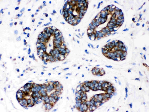
Figure 4. IHC analysis of CYP1A1 using anti-CYP1A1 antibody (MBS178240).
CYP1A1 was detected in paraffin-embedded section of Human Mammary Cancer Tissue. Heat mediated antigen retrieval was performed in citrate buffer (pH6, epitope retrieval solution) for 20 mins. The tissue section was blocked with 10% goat serum. The tissue section was then incubated with 1ug/ml rabbit anti-CYP1A1 Antibody (MBS178240) overnight at 4 degree C. Biotinylated goat anti-rabbit IgG was used as secondary antibody and incubated for 30 minutes at 37 degree C. The tissue section was developed using Strepavidin-Biotin-Complex (SABC) with DAB as the chromogen.
CYP1A1 was detected in paraffin-embedded section of Human Mammary Cancer Tissue. Heat mediated antigen retrieval was performed in citrate buffer (pH6, epitope retrieval solution) for 20 mins. The tissue section was blocked with 10% goat serum. The tissue section was then incubated with 1ug/ml rabbit anti-CYP1A1 Antibody (MBS178240) overnight at 4 degree C. Biotinylated goat anti-rabbit IgG was used as secondary antibody and incubated for 30 minutes at 37 degree C. The tissue section was developed using Strepavidin-Biotin-Complex (SABC) with DAB as the chromogen.
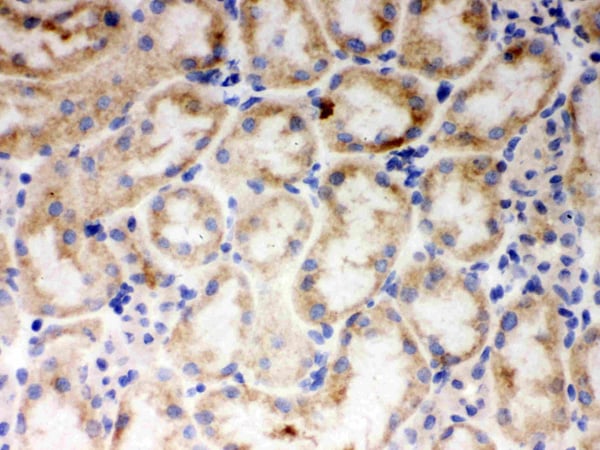
Figure 5. IHC analysis of CYP1A1 using anti-CYP1A1 antibody (MBS178240).
CYP1A1 was detected in frozen section of Mouse Kidney Tissue. Heat mediated antigen retrieval was performed in citrate buffer (pH6, epitope retrieval solution) for 20 mins. The tissue section was blocked with 10% goat serum. The tissue section was then incubated with 1ug/ml rabbit anti-CYP1A1 Antibody (MBS178240) overnight at 4 degree C. Biotinylated goat anti-rabbit IgG was used as secondary antibody and incubated for 30 minutes at 37 degree C. The tissue section was developed using Strepavidin-Biotin-Complex (SABC) with DAB as the chromogen.
CYP1A1 was detected in frozen section of Mouse Kidney Tissue. Heat mediated antigen retrieval was performed in citrate buffer (pH6, epitope retrieval solution) for 20 mins. The tissue section was blocked with 10% goat serum. The tissue section was then incubated with 1ug/ml rabbit anti-CYP1A1 Antibody (MBS178240) overnight at 4 degree C. Biotinylated goat anti-rabbit IgG was used as secondary antibody and incubated for 30 minutes at 37 degree C. The tissue section was developed using Strepavidin-Biotin-Complex (SABC) with DAB as the chromogen.
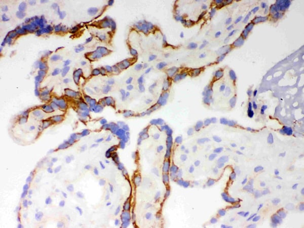
Figure 6. IHC analysis of CYP1A1 using anti-CYP1A1 antibody (MBS178240).
CYP1A1 was detected in frozen section of Human Placenta Tissue. Heat mediated antigen retrieval was performed in citrate buffer (pH6, epitope retrieval solution) for 20 mins. The tissue section was blocked with 10% goat serum. The tissue section was then incubated with 1ug/ml rabbit anti-CYP1A1 Antibody (MBS178240) overnight at 4 degree C. Biotinylated goat anti-rabbit IgG was used as secondary antibody and incubated for 30 minutes at 37 degree C. The tissue section was developed using Strepavidin-Biotin-Complex (SABC) with DAB as the chromogen.
CYP1A1 was detected in frozen section of Human Placenta Tissue. Heat mediated antigen retrieval was performed in citrate buffer (pH6, epitope retrieval solution) for 20 mins. The tissue section was blocked with 10% goat serum. The tissue section was then incubated with 1ug/ml rabbit anti-CYP1A1 Antibody (MBS178240) overnight at 4 degree C. Biotinylated goat anti-rabbit IgG was used as secondary antibody and incubated for 30 minutes at 37 degree C. The tissue section was developed using Strepavidin-Biotin-Complex (SABC) with DAB as the chromogen.
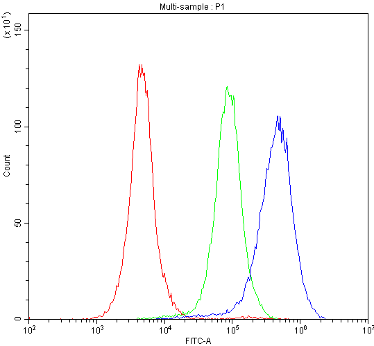
Figure 7. Flow Cytometry analysis of CACO-2 cells using anti-CYP1A1 antibody (MBS178240).
Overlay histogram showing CACO-2 cells stained with MBS178240 (Blue line).The cells were blocked with 10% normal goat serum. And then incubated with rabbit anti-CYP1A1 Antibody (MBS178240,1ug/1x10^6 cells) for 30 min at 20 degree C. DyLight®488 conjugated goat anti-rabbit IgG (5-10ug/1x10^6 cells) was used as secondary antibody for 30 minutes at 20 degree C. Isotype control antibody (Green line) was rabbit IgG (1ug/1x106) used under the same conditions. Unlabelled sample (Red line) was also used as a control.
Overlay histogram showing CACO-2 cells stained with MBS178240 (Blue line).The cells were blocked with 10% normal goat serum. And then incubated with rabbit anti-CYP1A1 Antibody (MBS178240,1ug/1x10^6 cells) for 30 min at 20 degree C. DyLight®488 conjugated goat anti-rabbit IgG (5-10ug/1x10^6 cells) was used as secondary antibody for 30 minutes at 20 degree C. Isotype control antibody (Green line) was rabbit IgG (1ug/1x106) used under the same conditions. Unlabelled sample (Red line) was also used as a control.
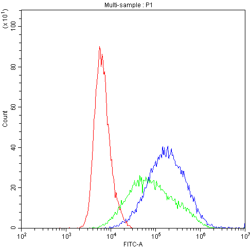
Figure 8. Flow Cytometry analysis of Hela cells using anti-CYP1A1 antibody (MBS178240).
Overlay histogram showing Hela cells stained with MBS178240 (Blue line).The cells were blocked with 10% normal goat serum. And then incubated with rabbit anti-CYP1A1 Antibody (MBS178240,1ug/1x10^6 cells) for 30 min at 20 degree C. DyLight®488 conjugated goat anti-rabbit IgG (5-10ug/1x10^6 cells) was used as secondary antibody for 30 minutes at 20 degree C. Isotype control antibody (Green line) was rabbit IgG (1ug/1x106) used under the same conditions. Unlabelled sample (Red line) was also used as a control.
Overlay histogram showing Hela cells stained with MBS178240 (Blue line).The cells were blocked with 10% normal goat serum. And then incubated with rabbit anti-CYP1A1 Antibody (MBS178240,1ug/1x10^6 cells) for 30 min at 20 degree C. DyLight®488 conjugated goat anti-rabbit IgG (5-10ug/1x10^6 cells) was used as secondary antibody for 30 minutes at 20 degree C. Isotype control antibody (Green line) was rabbit IgG (1ug/1x106) used under the same conditions. Unlabelled sample (Red line) was also used as a control.
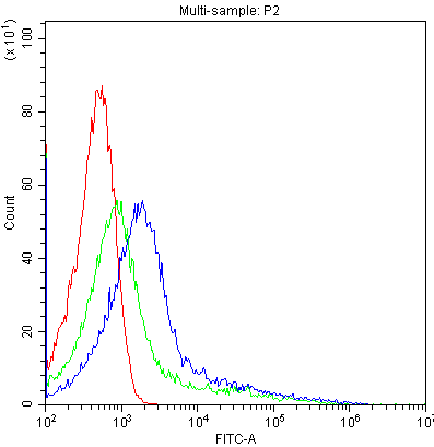
Figure 9. Flow Cytometry analysis of K562 cells using anti-CYP1A1 antibody (MBS178240).
Overlay histogram showing K562 cells stained with MBS178240 (Blue line).The cells were blocked with 10% normal goat serum. And then incubated with rabbit anti-CYP1A1 Antibody (MBS178240,1ug/1x10^6 cells) for 30 min at 20 degree C. DyLight®488 conjugated goat anti-rabbit IgG (5-10ug/1x10^6 cells) was used as secondary antibody for 30 minutes at 20 degree C. Isotype control antibody (Green line) was rabbit IgG (1ug/1x106) used under the same conditions. Unlabelled sample (Red line) was also used as a control.
Overlay histogram showing K562 cells stained with MBS178240 (Blue line).The cells were blocked with 10% normal goat serum. And then incubated with rabbit anti-CYP1A1 Antibody (MBS178240,1ug/1x10^6 cells) for 30 min at 20 degree C. DyLight®488 conjugated goat anti-rabbit IgG (5-10ug/1x10^6 cells) was used as secondary antibody for 30 minutes at 20 degree C. Isotype control antibody (Green line) was rabbit IgG (1ug/1x106) used under the same conditions. Unlabelled sample (Red line) was also used as a control.
CYP1A1 Antibody – Rabbit Polyclonal
For Research Use Only. Not for use in diagnostic procedures. Catalog #: MBS178240CYP1A1 is involved in phase I xenobiotic and drug metabolism (one substrate of it is theophylline). It is inhibited by fluoroquinolones and macrolides and induced by aromatic hydrocarbons. CYP1A1 is also known as AHH (aryl hydrocarbon hydroxylase). It is… More details
Product Overview
Online Datasheet in PDFProduct Name
CYP1A1, Polyclonal Antibody
Full Product Name
Anti-CYP1A1 Antibody
Product Gene Name
anti-CYP1A1 antibody
Product Synonym Names
Cytochrome P450 1A1; AHH; AHRR; Aryl hydrocarbon hydroxylase; CP 11; CP11; CP1A1_HUMAN; CYP 1; CYP1; CYP1A1; CYPIA1; Cytochrome P1 450 dioxin inducible; Cytochrome P1-450; Cytochrome P1-450 dioxin-inducible; Cytochrome P450 1A1; Cytochrome P450 family 1 subfamily A polypeptide 1; Cytochrome P450 form 6; Cytochrome P450 subfamily I (aromatic compound inducible) polypeptide 1; Cytochrome P450 subfamily I aromatic compound inducible polypeptide 1; Cytochrome P450-C; Cytochrome P450-P1; Flavoprotein-linked monooxygenase; Microsomal monooxygenase; P1 450; P450 C; P450 form 6; P450 P1; P450DX; Xenobiotic monooxygenase; cytochrome P450, family 1, subfamily A, polypeptide 1
Clonality
Polyclonal
Isotype
IgG
Host
Rabbit
Species Reactivity
Human, Mouse, Rat
Form/Format
Lyophilized. Each vial contains 5mg BSA, 0.9mg NaCl, 0.2mg Na2HPO4, 0.05mg NaN3.
Purity/Purification
Immunogen Affinity Purified
Specificity
No cross reactivity with other proteins.
Preparation and Storage
Store at -20 degrees C for one year, at 4 degrees C for one month. Avoid repeated freezing and thawing.
OMIM
108330
Immunogen
E Coli-derived human CYP1A1 recombinant protein (Position: H183-D320). Human CYP1A1 shares 81.2% amino acid (aa) sequence identity with both mouse and rat CYP1A1.
Reconstitution
Add 0.2ml of distilled water will yield a concentration of 500ug/ml.
Relevant Detection Systems
It is recommended for Enhanced Chemiluminescent Kit with anti-Rabbit IgG (please inquire) for Western blot, and HRP Conjugated anti-Rabbit IgG Super Vision Assay Kit (please inquire) for IHC(P), IHC(F) and ICC.
ISO Certification
Manufactured in an ISO 13485:2003 and EN ISO 13485:2012 Certified Laboratory.
Applications Tested/Suitable for anti-CYP1A1 antibody
Western Blot (WB), Immunohistochemistry-Paraffin (IHC-P), Immunohistochemistry-Frozen (IHC-F), Immunocytochemistry (ICC), Flow Cytometry (FC)
Application Notes for anti-CYP1A1 antibody
WB: Concentration: 0.1-0.5ug/ml; Tested Species: Human Mouse, Rat
IHC-P: Concentration: 0.5-1ug/ml; Tested Species: Human, Mouse, Rat; Antigen Retrieval: By Heat
IHC-F: Concentration: 0.5-1ug/ml; Tested Species: Human, Mouse
ICC/IF: Concentration: 2 ug/ml; Tested Species: Human
FC: Concentration: 1-3ug/1x106 cells; Tested Species: Human.
Tested Species: In house tested species with positive results.
By heat: Boiling the paraffin section 10mM citrate buffer, pH6.0 for 20mins is required for the staining of formalin/paraffin sections.
Other applications have not been tested.
Optimal dilutions should be determined by end user.
Western Blot (WB) of anti-CYP1A1 antibody
Figure 1. Western blot analysis of CYP1A1 using anti-CYP1A1 antibody (MBS178240).
Electrophoresis was performed on a 5-20% SDS-PAGE gel at 70V (Stacking gel) / 90V (Resolving gel) for 2-3 hours. The sample well of each lane was loaded with 50ug of sample under reducing conditions.
Lane 1: Rat Lung Tissue Lysate
Lane 2: Mouse Lung Tissue Lysate
Lane 3: Human Placenta Tissue Lysate
Lane 4: JURKAT Whole Cell Lysate
After Electrophoresis, proteins were transferred to a Nitrocellulose membrane at 150mA for 50-90 minutes. Blocked the membrane with 5% Non-fat Milk/ TBS for 1.5 hour at RT. The membrane was incubated with rabbit anti-CYP1A1 antigen affinity purified polyclonal antibody at 0.5ug/mL overnight at 4 degree C, then washed with TBS-0.1%Tween 3 times with 5 minutes each and probed with a goat anti-rabbit IgG-HRP secondary antibody at a dilution of 1:10000 for 1.5 hour at RT. The signal is developed using an Enhanced Chemiluminescent detection (ECL) kit with Tanon 5200 system. A specific band was detected for CYP1A1 at approximately 58KD. The expected band size for CYP1A1 is at 58KD.
Electrophoresis was performed on a 5-20% SDS-PAGE gel at 70V (Stacking gel) / 90V (Resolving gel) for 2-3 hours. The sample well of each lane was loaded with 50ug of sample under reducing conditions.
Lane 1: Rat Lung Tissue Lysate
Lane 2: Mouse Lung Tissue Lysate
Lane 3: Human Placenta Tissue Lysate
Lane 4: JURKAT Whole Cell Lysate
After Electrophoresis, proteins were transferred to a Nitrocellulose membrane at 150mA for 50-90 minutes. Blocked the membrane with 5% Non-fat Milk/ TBS for 1.5 hour at RT. The membrane was incubated with rabbit anti-CYP1A1 antigen affinity purified polyclonal antibody at 0.5ug/mL overnight at 4 degree C, then washed with TBS-0.1%Tween 3 times with 5 minutes each and probed with a goat anti-rabbit IgG-HRP secondary antibody at a dilution of 1:10000 for 1.5 hour at RT. The signal is developed using an Enhanced Chemiluminescent detection (ECL) kit with Tanon 5200 system. A specific band was detected for CYP1A1 at approximately 58KD. The expected band size for CYP1A1 is at 58KD.
Immunohistochemistry (IHC) of anti-CYP1A1 antibody
Figure 2. IHC analysis of CYP1A1 using anti-CYP1A1 antibody (MBS178240).
CYP1A1 was detected in paraffin-embedded section of Mouse Kidney Tissue. Heat mediated antigen retrieval was performed in citrate buffer (pH6, epitope retrieval solution) for 20 mins. The tissue section was blocked with 10% goat serum. The tissue section was then incubated with 1ug/ml rabbit anti-CYP1A1 Antibody (MBS178240) overnight at 4 degree C. Biotinylated goat anti-rabbit IgG was used as secondary antibody and incubated for 30 minutes at 37 degree C. The tissue section was developed using Strepavidin-Biotin-Complex (SABC) with DAB as the chromogen.
CYP1A1 was detected in paraffin-embedded section of Mouse Kidney Tissue. Heat mediated antigen retrieval was performed in citrate buffer (pH6, epitope retrieval solution) for 20 mins. The tissue section was blocked with 10% goat serum. The tissue section was then incubated with 1ug/ml rabbit anti-CYP1A1 Antibody (MBS178240) overnight at 4 degree C. Biotinylated goat anti-rabbit IgG was used as secondary antibody and incubated for 30 minutes at 37 degree C. The tissue section was developed using Strepavidin-Biotin-Complex (SABC) with DAB as the chromogen.
Immunohistochemistry (IHC) of anti-CYP1A1 antibody
Figure 3. IHC analysis of CYP1A1 using anti-CYP1A1 antibody (MBS178240).
CYP1A1 was detected in paraffin-embedded section of Rat Kidney Tissue. Heat mediated antigen retrieval was performed in citrate buffer (pH6, epitope retrieval solution) for 20 mins. The tissue section was blocked with 10% goat serum. The tissue section was then incubated with 1ug/ml rabbit anti-CYP1A1 Antibody (MBS178240) overnight at 4 degree C. Biotinylated goat anti-rabbit IgG was used as secondary antibody and incubated for 30 minutes at 37 degree C. The tissue section was developed using Strepavidin-Biotin-Complex (SABC) with DAB as the chromogen.
CYP1A1 was detected in paraffin-embedded section of Rat Kidney Tissue. Heat mediated antigen retrieval was performed in citrate buffer (pH6, epitope retrieval solution) for 20 mins. The tissue section was blocked with 10% goat serum. The tissue section was then incubated with 1ug/ml rabbit anti-CYP1A1 Antibody (MBS178240) overnight at 4 degree C. Biotinylated goat anti-rabbit IgG was used as secondary antibody and incubated for 30 minutes at 37 degree C. The tissue section was developed using Strepavidin-Biotin-Complex (SABC) with DAB as the chromogen.
Immunohistochemistry (IHC) of anti-CYP1A1 antibody
Figure 4. IHC analysis of CYP1A1 using anti-CYP1A1 antibody (MBS178240).
CYP1A1 was detected in paraffin-embedded section of Human Mammary Cancer Tissue. Heat mediated antigen retrieval was performed in citrate buffer (pH6, epitope retrieval solution) for 20 mins. The tissue section was blocked with 10% goat serum. The tissue section was then incubated with 1ug/ml rabbit anti-CYP1A1 Antibody (MBS178240) overnight at 4 degree C. Biotinylated goat anti-rabbit IgG was used as secondary antibody and incubated for 30 minutes at 37 degree C. The tissue section was developed using Strepavidin-Biotin-Complex (SABC) with DAB as the chromogen.
CYP1A1 was detected in paraffin-embedded section of Human Mammary Cancer Tissue. Heat mediated antigen retrieval was performed in citrate buffer (pH6, epitope retrieval solution) for 20 mins. The tissue section was blocked with 10% goat serum. The tissue section was then incubated with 1ug/ml rabbit anti-CYP1A1 Antibody (MBS178240) overnight at 4 degree C. Biotinylated goat anti-rabbit IgG was used as secondary antibody and incubated for 30 minutes at 37 degree C. The tissue section was developed using Strepavidin-Biotin-Complex (SABC) with DAB as the chromogen.
Immunohistochemistry (IHC) of anti-CYP1A1 antibody
Figure 5. IHC analysis of CYP1A1 using anti-CYP1A1 antibody (MBS178240).
CYP1A1 was detected in frozen section of Mouse Kidney Tissue. Heat mediated antigen retrieval was performed in citrate buffer (pH6, epitope retrieval solution) for 20 mins. The tissue section was blocked with 10% goat serum. The tissue section was then incubated with 1ug/ml rabbit anti-CYP1A1 Antibody (MBS178240) overnight at 4 degree C. Biotinylated goat anti-rabbit IgG was used as secondary antibody and incubated for 30 minutes at 37 degree C. The tissue section was developed using Strepavidin-Biotin-Complex (SABC) with DAB as the chromogen.
CYP1A1 was detected in frozen section of Mouse Kidney Tissue. Heat mediated antigen retrieval was performed in citrate buffer (pH6, epitope retrieval solution) for 20 mins. The tissue section was blocked with 10% goat serum. The tissue section was then incubated with 1ug/ml rabbit anti-CYP1A1 Antibody (MBS178240) overnight at 4 degree C. Biotinylated goat anti-rabbit IgG was used as secondary antibody and incubated for 30 minutes at 37 degree C. The tissue section was developed using Strepavidin-Biotin-Complex (SABC) with DAB as the chromogen.
Immunohistochemistry (IHC) of anti-CYP1A1 antibody
Figure 6. IHC analysis of CYP1A1 using anti-CYP1A1 antibody (MBS178240).
CYP1A1 was detected in frozen section of Human Placenta Tissue. Heat mediated antigen retrieval was performed in citrate buffer (pH6, epitope retrieval solution) for 20 mins. The tissue section was blocked with 10% goat serum. The tissue section was then incubated with 1ug/ml rabbit anti-CYP1A1 Antibody (MBS178240) overnight at 4 degree C. Biotinylated goat anti-rabbit IgG was used as secondary antibody and incubated for 30 minutes at 37 degree C. The tissue section was developed using Strepavidin-Biotin-Complex (SABC) with DAB as the chromogen.
CYP1A1 was detected in frozen section of Human Placenta Tissue. Heat mediated antigen retrieval was performed in citrate buffer (pH6, epitope retrieval solution) for 20 mins. The tissue section was blocked with 10% goat serum. The tissue section was then incubated with 1ug/ml rabbit anti-CYP1A1 Antibody (MBS178240) overnight at 4 degree C. Biotinylated goat anti-rabbit IgG was used as secondary antibody and incubated for 30 minutes at 37 degree C. The tissue section was developed using Strepavidin-Biotin-Complex (SABC) with DAB as the chromogen.
Flow Cytometry (FC/FACS) of anti-CYP1A1 antibody
Figure 7. Flow Cytometry analysis of CACO-2 cells using anti-CYP1A1 antibody (MBS178240).
Overlay histogram showing CACO-2 cells stained with MBS178240 (Blue line).The cells were blocked with 10% normal goat serum. And then incubated with rabbit anti-CYP1A1 Antibody (MBS178240,1ug/1x10^6 cells) for 30 min at 20 degree C. DyLight®488 conjugated goat anti-rabbit IgG (5-10ug/1x10^6 cells) was used as secondary antibody for 30 minutes at 20 degree C. Isotype control antibody (Green line) was rabbit IgG (1ug/1x106) used under the same conditions. Unlabelled sample (Red line) was also used as a control.
Overlay histogram showing CACO-2 cells stained with MBS178240 (Blue line).The cells were blocked with 10% normal goat serum. And then incubated with rabbit anti-CYP1A1 Antibody (MBS178240,1ug/1x10^6 cells) for 30 min at 20 degree C. DyLight®488 conjugated goat anti-rabbit IgG (5-10ug/1x10^6 cells) was used as secondary antibody for 30 minutes at 20 degree C. Isotype control antibody (Green line) was rabbit IgG (1ug/1x106) used under the same conditions. Unlabelled sample (Red line) was also used as a control.
Flow Cytometry (FC/FACS) of anti-CYP1A1 antibody
Figure 8. Flow Cytometry analysis of Hela cells using anti-CYP1A1 antibody (MBS178240).
Overlay histogram showing Hela cells stained with MBS178240 (Blue line).The cells were blocked with 10% normal goat serum. And then incubated with rabbit anti-CYP1A1 Antibody (MBS178240,1ug/1x10^6 cells) for 30 min at 20 degree C. DyLight®488 conjugated goat anti-rabbit IgG (5-10ug/1x10^6 cells) was used as secondary antibody for 30 minutes at 20 degree C. Isotype control antibody (Green line) was rabbit IgG (1ug/1x106) used under the same conditions. Unlabelled sample (Red line) was also used as a control.
Overlay histogram showing Hela cells stained with MBS178240 (Blue line).The cells were blocked with 10% normal goat serum. And then incubated with rabbit anti-CYP1A1 Antibody (MBS178240,1ug/1x10^6 cells) for 30 min at 20 degree C. DyLight®488 conjugated goat anti-rabbit IgG (5-10ug/1x10^6 cells) was used as secondary antibody for 30 minutes at 20 degree C. Isotype control antibody (Green line) was rabbit IgG (1ug/1x106) used under the same conditions. Unlabelled sample (Red line) was also used as a control.
Flow Cytometry (FC/FACS) of anti-CYP1A1 antibody
Figure 9. Flow Cytometry analysis of K562 cells using anti-CYP1A1 antibody (MBS178240).
Overlay histogram showing K562 cells stained with MBS178240 (Blue line).The cells were blocked with 10% normal goat serum. And then incubated with rabbit anti-CYP1A1 Antibody (MBS178240,1ug/1x10^6 cells) for 30 min at 20 degree C. DyLight®488 conjugated goat anti-rabbit IgG (5-10ug/1x10^6 cells) was used as secondary antibody for 30 minutes at 20 degree C. Isotype control antibody (Green line) was rabbit IgG (1ug/1x106) used under the same conditions. Unlabelled sample (Red line) was also used as a control.
Overlay histogram showing K562 cells stained with MBS178240 (Blue line).The cells were blocked with 10% normal goat serum. And then incubated with rabbit anti-CYP1A1 Antibody (MBS178240,1ug/1x10^6 cells) for 30 min at 20 degree C. DyLight®488 conjugated goat anti-rabbit IgG (5-10ug/1x10^6 cells) was used as secondary antibody for 30 minutes at 20 degree C. Isotype control antibody (Green line) was rabbit IgG (1ug/1x106) used under the same conditions. Unlabelled sample (Red line) was also used as a control.
Product References and Citations for anti-CYP1A1 antibody
1. Beresford AP (1993). "CYP1A1: friend or foe?". Drug Metabolism Reviews 25 (4): 503-17. 2. Uno, S (2004). "Oral exposure to benzoampyrene in the mouse: detoxication by inducible cytochrome P450 is more important than metabolic activation.". Mol Pharmacol 65 (5): 1225-37.
Other Notes
Small volumes of anti-CYP1A1 antibody vial(s) may occasionally become entrapped in the seal of the product vial during shipment and storage. If necessary, briefly centrifuge the vial on a tabletop centrifuge to dislodge any liquid in the container`s cap. Certain products may require to ship with dry ice and additional dry ice fee may apply.
Searchable Terms for CYP1A1 purchase
MBS178240 is an antibody targeting CYP1A1. Reactive in Human, Mouse, Rat. Host: Rabbit.
Additional Details
NCBI/Uniprot data below describe general gene information for CYP1A1. It may not necessarily be applicable to this product.
NCBI Official Full Name
cytochrome P450 1A1 isoform 1
NCBI Official Synonym Full Names
cytochrome P450 family 1 subfamily A member 1
NCBI Official Symbol
CYP1A1
NCBI Official Synonym Symbols
AHH; AHRR; CP11; CYP1; CYPIA1; P1-450; P450-C; P450DX
UniProt Protein Name
Cytochrome P450 1A1
UniProt Synonym Protein Names
CYPIA1; Cytochrome P450 form 6; Cytochrome P450-C; Cytochrome P450-P1
Protein Family
Cytochrome
UniProt Gene Name
CYP1A1
UniProt Entry Name
CP1A1_HUMAN
NCBI Protein Information
cytochrome P450 1A1
NCBI Summary for CYP1A1
This gene, CYP1A1, encodes a member of the cytochrome P450 superfamily of enzymes. The cytochrome P450 proteins are monooxygenases which catalyze many reactions involved in drug metabolism and synthesis of cholesterol, steroids and other lipids. This protein localizes to the endoplasmic reticulum and its expression is induced by some polycyclic aromatic hydrocarbons (PAHs), some of which are found in cigarette smoke. The enzyme's endogenous substrate is unknown; however, it is able to metabolize some PAHs to carcinogenic intermediates. The gene has been associated with lung cancer risk. A related family member, CYP1A2, is located approximately 25 kb away from CYP1A1 on chromosome 15. Alternative splicing results in multiple transcript variants encoding distinct isoforms. [provided by RefSeq, Jan 2016]
Show more UniProt Comments for CYP1A1
CYP1A1: Cytochromes P450 are a group of heme-thiolate monooxygenases. In liver microsomes, this enzyme is involved in an NADPH-dependent electron transport pathway. It oxidizes a variety of structurally unrelated compounds, including steroids, fatty acids, and xenobiotics. Belongs to the cytochrome P450 family.
Protein type: Oxidoreductase; Cofactor and Vitamin Metabolism - retinol; Amino Acid Metabolism - tryptophan; Xenobiotic Metabolism - metabolism by cytochrome P450; EC 1.14.14.1
Chromosomal Location of Human Ortholog: 15q24.1
Cellular Component: endoplasmic reticulum membrane; intracellular membrane-bound organelle; mitochondrion
Molecular Function: demethylase activity; enzyme binding; flavonoid 3'-monooxygenase activity; heme binding; iron ion binding; monooxygenase activity; oxidoreductase activity; oxidoreductase activity, acting on diphenols and related substances as donors; oxygen binding; protein binding; steroid hydroxylase activity
Biological Process: 9-cis-retinoic acid biosynthetic process; aging; amine metabolic process; camera-type eye development; cell proliferation; coumarin metabolic process; dibenzo-p-dioxin catabolic process; drug metabolic process; embryonic development ending in birth or egg hatching; epoxygenase P450 pathway; ethylene metabolic process; flavonoid metabolic process; gut development; hydrogen peroxide biosynthetic process; insecticide metabolic process; maternal process involved in parturition; porphyrin metabolic process; response to antibiotic; response to arsenic; response to drug; response to food; response to herbicide; response to hyperoxia; response to hypoxia; response to iron(III) ion; response to lipopolysaccharide; response to nematode; response to virus; response to vitamin A; response to wounding; vitamin D metabolic process
Show more Protein type: Oxidoreductase; Cofactor and Vitamin Metabolism - retinol; Amino Acid Metabolism - tryptophan; Xenobiotic Metabolism - metabolism by cytochrome P450; EC 1.14.14.1
Chromosomal Location of Human Ortholog: 15q24.1
Cellular Component: endoplasmic reticulum membrane; intracellular membrane-bound organelle; mitochondrion
Molecular Function: demethylase activity; enzyme binding; flavonoid 3'-monooxygenase activity; heme binding; iron ion binding; monooxygenase activity; oxidoreductase activity; oxidoreductase activity, acting on diphenols and related substances as donors; oxygen binding; protein binding; steroid hydroxylase activity
Biological Process: 9-cis-retinoic acid biosynthetic process; aging; amine metabolic process; camera-type eye development; cell proliferation; coumarin metabolic process; dibenzo-p-dioxin catabolic process; drug metabolic process; embryonic development ending in birth or egg hatching; epoxygenase P450 pathway; ethylene metabolic process; flavonoid metabolic process; gut development; hydrogen peroxide biosynthetic process; insecticide metabolic process; maternal process involved in parturition; porphyrin metabolic process; response to antibiotic; response to arsenic; response to drug; response to food; response to herbicide; response to hyperoxia; response to hypoxia; response to iron(III) ion; response to lipopolysaccharide; response to nematode; response to virus; response to vitamin A; response to wounding; vitamin D metabolic process
Precautions
All of MyBioSource's Products are for scientific laboratory research purposes and are not for diagnostic, therapeutics, prophylactic or in vivo use. Through your purchase, you expressly represent and warrant to MyBioSource that you will properly test and use any Products purchased from MyBioSource in accordance with industry standards. MyBioSource and its authorized distributors reserve the right to refuse to process any order where we reasonably believe that the intended use will fall outside of our acceptable guidelines.
Disclaimer
While every efforts were made to ensure the accuracy of the information provided in this datasheet, MyBioSource will not be liable for any omissions or errors contained herein. MyBioSource reserves the right to make changes to this datasheet at any time without prior notice.
It is the responsibility of the customer to report product performance issues to MyBioSource within 30 days of receipt of the product. Please visit our Terms & Conditions page for more information.
It is the responsibility of the customer to report product performance issues to MyBioSource within 30 days of receipt of the product. Please visit our Terms & Conditions page for more information.
Product Note
Select online data sheet information is drawn from bioinformatics databases, occasionally resulting in ambiguous or non-relevant product information. It is the responsibility of the customer to review, verify, and evaluate the information to make sure it matches their requirements before purchasing the kit. Our ELISA Kit assays are dynamic research tools and sometimes they may be updated and improved. If the format of this assay is important to you then please request the current manual or contact our technical support team with a presales inquiry before placing an order. We will confirm the current details of the assay. We cannot guarantee the sample manual posted online is the most current manual, it is intended to serve as an example only. Please refer to the instructions For Use provided with the assay kit for precise details.
Product PubMed Publications
Products associated with anti-CYP1A1 antibody
| Reference Product | PubMed Publications |
|---|---|
| AHR antibody | >1566 publications with CYP1A1 and AHR |
| GSTM1 antibody | >662 publications with CYP1A1 and GSTM1 |
| GSTT1 antibody | >429 publications with CYP1A1 and GSTT1 |
| GSTP1 antibody | >297 publications with CYP1A1 and GSTP1 |
| EPHX1 antibody | >191 publications with CYP1A1 and EPHX1 |
| COMT antibody | >120 publications with CYP1A1 and COMT |
| UGT1A1 antibody | >78 publications with CYP1A1 and UGT1A1 |
| UGT1A6 antibody | >52 publications with CYP1A1 and UGT1A6 |
| UGT1A7 antibody | >14 publications with CYP1A1 and UGT1A7 |
| UGT1A10 antibody | >3 publications with CYP1A1 and UGT1A10 |
Pathways associated with anti-CYP1A1 antibody
| Products by Pathway | Pathway Diagram |
|---|---|
| AhR Pathway antibodies | AhR Pathway Diagram |
| Arachidonic Acid Metabolism Pathway antibodies | Arachidonic Acid Metabolism Pathway Diagram |
| Benzo(a)pyrene Metabolism Pathway antibodies | Benzo(a)pyrene Metabolism Pathway Diagram |
| Biological Oxidations Pathway antibodies | Biological Oxidations Pathway Diagram |
| Chemical Carcinogenesis Pathway antibodies | Chemical Carcinogenesis Pathway Diagram |
| Chemical Carcinogenesis Pathway antibodies | Chemical Carcinogenesis Pathway Diagram |
| Cytochrome P450 - Arranged By Substrate Type Pathway antibodies | Cytochrome P450 - Arranged By Substrate Type Pathway Diagram |
| Estrogen Metabolism Pathway antibodies | Estrogen Metabolism Pathway Diagram |
| Fatty Acid Omega Oxidation Pathway antibodies | Fatty Acid Omega Oxidation Pathway Diagram |
| Fatty Acid, Triacylglycerol, And Ketone Body Metabolism Pathway antibodies | Fatty Acid, Triacylglycerol, And Ketone Body Metabolism Pathway Diagram |
Diseases associated with anti-CYP1A1 antibody
| Disease Name | Pubmed Publications |
|---|---|
| Liver Diseases Antibodies | >574 publications with CYP1A1 and Liver Diseases |
| Liver Neoplasms Antibodies | >507 publications with CYP1A1 and Liver Neoplasms |
| Lung Neoplasms Antibodies | >377 publications with CYP1A1 and Lung Neoplasms |
| Breast Neoplasms Antibodies | >374 publications with CYP1A1 and Breast Neoplasms |
| Neoplasms, Experimental Antibodies | >279 publications with CYP1A1 and Neoplasms, Experimental |
| Carcinoma, Hepatocellular Antibodies | >235 publications with CYP1A1 and Carcinoma, Hepatocellular |
| Inflammation Antibodies | >211 publications with CYP1A1 and Inflammation |
| Liver Neoplasms, Experimental Antibodies | >205 publications with CYP1A1 and Liver Neoplasms, Experimental |
| Necrosis Antibodies | >160 publications with CYP1A1 and Necrosis |
| Poisoning Antibodies | >113 publications with CYP1A1 and Poisoning |
Organs/Tissues associated with anti-CYP1A1 antibody
| Organ/Tissue Name | Pubmed Publications |
|---|---|
| Liver Antibodies | >3953 publications with CYP1A1 and Liver |
| Blood Antibodies | >1171 publications with CYP1A1 and Blood |
| Lung Antibodies | >923 publications with CYP1A1 and Lung |
| Brain Antibodies | >302 publications with CYP1A1 and Brain |
| Intestine Antibodies | >259 publications with CYP1A1 and Intestine |
| Skin Antibodies | >233 publications with CYP1A1 and Skin |
| Heart Antibodies | >174 publications with CYP1A1 and Heart |
| Embryonic Tissue Antibodies | >130 publications with CYP1A1 and Embryonic Tissue |
| Prostate Antibodies | >102 publications with CYP1A1 and Prostate |
| Uterus Antibodies | >86 publications with CYP1A1 and Uterus |
Sie könnten auch an folgenden Produkten interessiert sein:
Katalog-Nummer
Beschreibung
Cond.
Preis zzgl. MwSt.