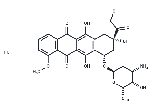
Doxorubicin hydrochloride [25316-40-9]

Katalog-Nummer T1020-25mg
Size : 25mg
Marke : TargetMol
Doxorubicin hydrochloride
(Synonyms: NSC 123127, Hydroxydaunorubicin hydrochloride, DOX hydrochloride, Adriamycin HCl) Copy Product Info

Product Introduction
| Description | Doxorubicin hydrochloride is an anthracycline antibiotic with cytotoxic and antitumor activity. It is an effective inhibitor of human DNA topoisomerase I and II, with IC50 values of 0.8 μM and 2.67 μM, respectively. In addition to inhibiting DNA topoisomerases, it can reduce the phosphorylation of AMPK and its downstream target, acetyl-CoA carboxylase. Doxorubicin hydrochloride also induces apoptosis and autophagy. In animal studies, it is commonly used to induce models of acute renal failure, chronic kidney injury, and heart failure. |
| Targets & IC50 | Topo II:2.67 μM, Topo I:0.8 μM |
| In vitro | METHODS: Human breast cancer cells MCF10A, BT474, MCF-7 and T47D were treated with Doxorubicin hydrochloride (0.1-10 μM) for 48 h, and cell growth inhibition was detected by MTT. RESULTS: Doxorubicin hydrochloride dose-dependently inhibited the growth of MCF10A, BT474, MCF-7 and T47D cells with IC50s of 2.51 µM, 1.14 µM, 0.69 µM and 8.53 µM, respectively. [1] METHODS: Bovine aortic endothelial cells BAECs and human ovarian teratoma cells PA-1 were treated with Doxorubicin (0.5 μM) for 1-16 h. Apoptosis was detected by Flow Cytometry, and caspase-3 activity was detected by caspase-3 assay kit. RESULTS: Doxorubicin induced apoptosis and caspase-3 activation in BAECs and PA-1 cells in a time-dependent manner. [2] METHODS: Canine breast cancer cells CIPp were treated with Doxorubicin (EC50(20h)=12.08 μM) for 3-48 h. The expression of target genes was detected by qRT-PCR. RESULTS: Doxorubicin induced up-regulation of the mRNA expression levels of multidrug resistance (MDR)-related genes P-gp and BCRP. [3] |
| In vivo | METHODS: To detect antitumor activity in vivo, Doxorubicin hydrochloride (1 mg/kg/4 days) and lovastatin (5 mg/kg/day) were intraperitoneally injected into B6D2F1 mice bearing murine melanoma tumor B16F10 for two weeks. RESULTS: The combination of Doxorubicin hydrochloride and lovastatin showed a significant increase in sensitivity compared to either drug alone. lovastatin enhanced the antitumor activity of Doxorubicin hydrochloride. [4] METHODS: To investigate the acute and long-term cognitive deficits of Doxorubicin in cancer patients, a single dose of Doxorubicin hydrochloride (25 mg/kg) was administered intraperitoneally to B6C3F1J mice. RESULTS: Systemic treatment with Doxorubicin hydrochloride altered glutamatergic neurotransmission in the nucleus of key cells associated with cognitive function within 24 h. There were no lasting effects on spatial learning and memory. [5] |
| Synonyms | NSC 123127, Hydroxydaunorubicin hydrochloride, DOX hydrochloride, Adriamycin HCl |
| Disease Modeling Protocol | Chronic kidney disease modeling
*Precautions: All rats were sacrificed 14 days after modeling, and plasma and kidney tissue were collected for testing. *References:Ikewuchi CC,et,al. Moderation of doxorubicin-induced nephrotoxicity in Wistar rats by aqueous leaf-extracts of Chromolaena odorata and Tridax procumbens. Porto Biomed J. 2021 Feb 11;6(1):e129. |
| Cell Research | To analyze the effect of Bcl-2 expression on the viability of HUVECs treated with Dox, cells were co-transfected with 200 ng of the pEGFP-spectrin expression plasmid together with 200 ng of either pCDNA3-hBcl-2 or the control pCMVβ-galactosidase expression vector (33). The pGL3 Basic vector (2.1 μg) was added as a DNA carrier in a total volume of 0.140 ml, and transfection was performed by the calcium phosphate procedure in 35-mm tissue culture dishes. After treatment, the cells were washed with PBS, fixed with 3.7% formaldehyde for 15 min, and washed for a further 10 min with 50 mM NH4Cl blocking solution in PBS. Cells were then washed with PBS, permeabilized with a 0.1% Triton X-100 for 10 min, washed again with PBS, and stained with 1 μg/ml 4′,6-diamidino-2-phenyl-indole solution for 2 min. The cells were examined under a fluorescence microscope, and GFP-positive cells were scored after counting a minimum of 1000 total cells for each condition. The efficiency of transfection in Bcl-2- and β-galactosidase-expressing cells, determined in aliquots of transfected cells just before the addition of Dox, was similar (10–12%) [1]. |
| Animal Research | Athymic male nude mice (3-4 weeks old) are used. PC3 cells (4×10⁶) are injected subcutaneously into the flanks of mice. Animals bearing tumors are randomly assigned to treatment groups (five or six mice per group) and treatment initiated when xenografts reached volumes of about 100 mm3. Tumors are measured using digital calipers and volume calculated using the formula: Volume=Width2×Length×0.52, where width represents the shorter dimension of the tumor. Treatments are administered as indicated using vehicle (PBS containing 0.1% BSA), Doxorubicin (2-8 mg/kg), Apo2L/TRAIL (500 μg/animal), or a combination of 4 mg/kg Doxorubicin followed by 500 μg Apo2L/TRAIL. Doxorubicin is administered systemically whereas Apo2L/TRAIL is given either intratumorally or systemically. All treatments are given once. Mice are monitored daily for signs of adverse effects (listlessness and scruffy appearance). Treatments seemed to be well tolerated. The mean±SEM is calculated for each data point. Differences between treatment groups are analyzed by the student t-test. Differences are considered significant when P<0.05 [3]. Altogether, 29 male Wistar rats (weight 306 ± 18.6 g) were used in the study. Animals were divided into three groups: control (group C; n = 10; 306.4 ± 17.2 g), animals treated with DOX (group DOX; n = 10; 305.0 ± 24.9 g) and animals treated with L-DOX (group L-DOX; n = 9; 306.7 ± 15.0 g). Vehiculum (aqua pro injection), DOX and L-DOX were applied to group C, DOX and L-DOX, respectively, by single intraperitoneal injection; concentration of both DOX and L-DOX was 5 mg/kg, similar to the concentrations used in human treatment protocols. All animals were sacrificed 24 h after drug application. Thoracotomy was performed, hearts were excised and samples were obtained separately from the free wall of the left atrium (LA), left ventricle (LV), right atrium (RA) and right ventricle (RV).Samples were placed into RNA later preservation solution and stored at -80 C until further analysis [4]. |
| Molecular Weight | 579.99 |
| Formula | C27H29NO11·HCl |
| Cas No. | 25316-40-9 |
| Smiles | Cl.COc1cccc2C(=O)c3c(O)c4C[C@](O)(C[C@H](O[C@H]5C[C@H](N)[C@H](O)[C@H](C)O5)c4c(O)c3C(=O)c12)C(=O)CO |
| Relative Density. | no data available |
| Storage | Keep away from direct sunlight,Keep away from moisture Powder: -20°C for 3 years | In solvent: -80°C for 1 year Shipping with blue ice/Shipping at ambient temperature. | ||||||||||||||||||||||||||||||||||||||||
| Solubility Information | DMSO: 240 mg/mL (413.8 mM), Sonication is recommended.  H2O: 50 mg/mL (86.21 mM), Sonication is recommended. | ||||||||||||||||||||||||||||||||||||||||
| In Vivo Formulation | 10% DMSO+40% PEG300+5% Tween 80+45% Saline: 5 mg/mL (8.62 mM), Sonication is recommended. Please add the solvents sequentially, clarifying the solution as much as possible before adding the next one. Dissolve by heating and/or sonication if necessary. Working solution is recommended to be prepared and used immediately. The formulation provided above is for reference purposes only. In vivo formulations may vary and should be modified based on specific experimental conditions. | ||||||||||||||||||||||||||||||||||||||||
Solution Preparation Table | |||||||||||||||||||||||||||||||||||||||||
H2O/DMSO
DMSO
Note : The dilution table applies only to solid products. For liquid products, please calculate the stock solution based on the stated concentration and/or density. | |||||||||||||||||||||||||||||||||||||||||