

NucView® 488 and RedDot™ 2 Apoptosis & Necrosis Kit (100 assays)

Katalog-Nummer 30072
Size : 1kit
Marke : Biotium
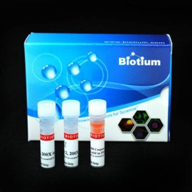
NucView® 488 & RedDot™ 2 Apoptosis & Necrosis Kit
Homogeneous, no wash assay for detection of caspase-3/7 activity with green fluorescence and dead cell staining with far-red fluorescence, for fluorescence microscopy or flow cytometry.
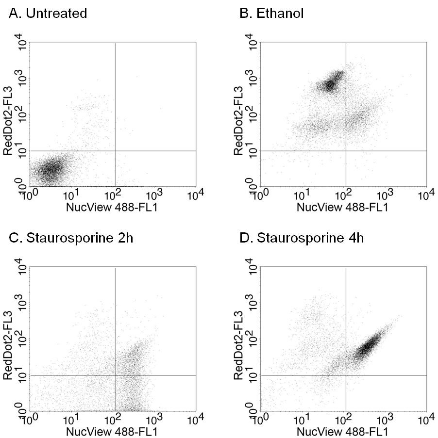 Flow cytometry analysis of Jurkat cells left untreated (A), treated with 10%
ethanol for 90 minutes to induce necrosis (B), or treated with 1 uM staurosporine for
2 hours (C) or 4 hours (D) to induce apoptosis. Cells were stained for 30 minutes at
room temperature with 2.5 uM NucView® 488 and 0.25X RedDot™2. NucView® 488
was detected in the FL1 channel (488 nm excitation and 530/30 nm emission filter) and
RedDot™2 in the FL3 channel (488 nm excitation/670 longpass emission filter) of a BD
FACSCalibur flow cytometer. Necrotic cells stain low with NucView® 488 and high with
RedDot™2. Early apoptotic cells stain high with NucView® 488 and low with RedDot™2,
while late apoptotic cells stain high with both NucView® 488 and RedDot™2.
Flow cytometry analysis of Jurkat cells left untreated (A), treated with 10%
ethanol for 90 minutes to induce necrosis (B), or treated with 1 uM staurosporine for
2 hours (C) or 4 hours (D) to induce apoptosis. Cells were stained for 30 minutes at
room temperature with 2.5 uM NucView® 488 and 0.25X RedDot™2. NucView® 488
was detected in the FL1 channel (488 nm excitation and 530/30 nm emission filter) and
RedDot™2 in the FL3 channel (488 nm excitation/670 longpass emission filter) of a BD
FACSCalibur flow cytometer. Necrotic cells stain low with NucView® 488 and high with
RedDot™2. Early apoptotic cells stain high with NucView® 488 and low with RedDot™2,
while late apoptotic cells stain high with both NucView® 488 and RedDot™2.Product Description
The NucView® 488 and RedDot™ Apoptosis & Necrosis Assay Kit provides a convenient tool for profiling apoptotic and necrotic cell populations by fluorescence microscopy or flow cytometry.
- Rapid, no-wash, endpoint or real-time assay
- Non-toxic, allowing multi-day experiments to be performed
- For flow cytometry, microscopy or live cell imaging systems
- Dual detection of caspase activity (green) and necrotic cells (far-red)
This kit contains NucView® 488 Caspase-3 Substrate for detection of caspase-3/7 activity. The far-red dead cell stain RedDot™ is included for staining necrotic and late apoptotic cells that have compromised plasma membrane integrity.
In contrast to other fluorogenic caspase substrates or fluorescent caspase inhibitor based (FLICA) assays, NucView® 488 Caspase-3 Substrate can be used to detect caspase-3/7 activity within individual intact cells without inhibiting apoptosis progression. The substrate consists of a fluorogenic DNA dye coupled to the caspase-3/7 DEVD recognition sequence. The substrate, which is initially non-fluorescent, penetrates the plasma membrane and enters the cytoplasm. In apoptotic cells, caspase-3/7 cleaves the substrate, releasing the high-affinity DNA dye, which migrates to the cell nucleus and stains DNA with bright green fluorescence. Thus, NucView® 488 Caspase-3 Substrate allows detection caspase-3/7 activity and visualization of morphological changes in the nucleus during apoptosis.
RedDot™2 is a cell membrane-impermeable, far-red dye with high selectivity for membrane compromised or dead cells. RedDot™2 has far-red emission at 695 nm for detection in the Cy®5 channel, well-separated from the green fluorescence of NucView® 488. The excitation maximum of RedDot™2 is 665 nm, but it can be efficiently excited by wavelengths from 488 to 647 nm, and therefore can be used with the 488 nm flow cytometry laser line.
Note: While NucView® 488 staining is formaldehyde-fixable, fixation after staining with RedDot™2 is not recommended because it can result in increased background staining of healthy cells.
To learn about the advantages of monitoring apoptosis using NucView® caspase-3 substrates, visit the NucView® Technology Page.
Technology

The Full Spectrum Blog

Product Attributes
Size
Apoptosis/viability marker
For live or fixed cells
Detection method/readout
Assay type/options
Substrate specificity
Colors
Storage Conditions
Documents, Protocols, SDS and COA

COA
Request the Certificate of AnalysisReferences
1. Cytometry Part A (2014) 85A, 179187. DOI: 10.1002/cyto.a.22410
2. bioRxiv (2019) doi: https://doi.org/10.1101/542597
3. Cellular Microbiol (2019) 21, e12956. https://doi.org/10.1111/cmi.12956
4. Toxicol Rep (2019) 6, 305-320. https://doi.org/10.1016/j.toxrep.2019.02.004
Find a list of NucView® references and a list of validated cell lines under Supporting Documents.
Citations
1. Cytometry Part A (2014) 85A, 179187. DOI: 10.1002/cyto.a.22410
2. bioRxiv (2019) doi: https://doi.org/10.1101/542597
3. Cellular Microbiol (2019) 21, e12956. https://doi.org/10.1111/cmi.12956
4. Toxicol Rep (2019) 6, 305-320. https://doi.org/10.1016/j.toxrep.2019.02.004
Find a list of NucView® references and a list of validated cell lines under Supporting Documents.
 Powered by Bioz