

ViaFluor® 488 SE Cell Proliferation Kit (10 vials, 1000 assays)

Katalog-Nummer 30086
Size : 1kit
Marke : Biotium
ViaFluor® SE Cell Proliferation Kits
Fixable cytoplasmic stains for monitoring cell division by flow cytometry. The dyes can also be used to track cell populations in co-culture.
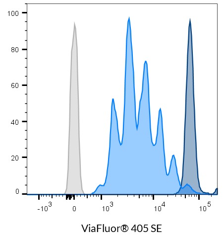 PBMCs were stained with 5 uM ViaFluor 405 Cell Proliferation Dye, and then T cell proliferation was stimulated with CD3/CD28 T Cell Activator Beads and IL-2. After 4 days, the cells were analyzed by flow cytometry in the Pacific Blue channel. The stimulated T cell proliferation can be measured by the number of individual peaks (light blue). Unstimulated cells are shown in dark blue, and unstained cells are in gray.
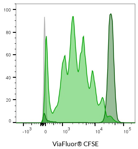 PBMCs were stained with 1 uM ViaFluor CFSE Cell Proliferation Dye, and then T cell proliferation was stimulated with CD3/CD28 T Cell Activator Beads and IL-2. After 4 days, the cells were analyzed by flow cytometry in the B525 (FITC) channel. The stimulated T cell proliferation can be measured by the number of individual peaks (light green). Unstimulated cells are shown in dark green, and unstained cells and in gray.
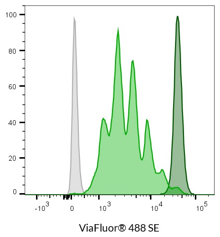 PBMCs were stained with 1 uM ViaFluor 488 Cell Proliferation Dye, and then T cell proliferation was stimulated with CD3/CD28 T Cell Activator Beads and IL-2. After 4 days, the cells were analyzed by flow cytometry in the B525 (FITC) channel. The stimulated T cell proliferation can be measured by the number of individual peaks (light green). Unstimulated cells are shown in dark green, and unstained cells and in gray.
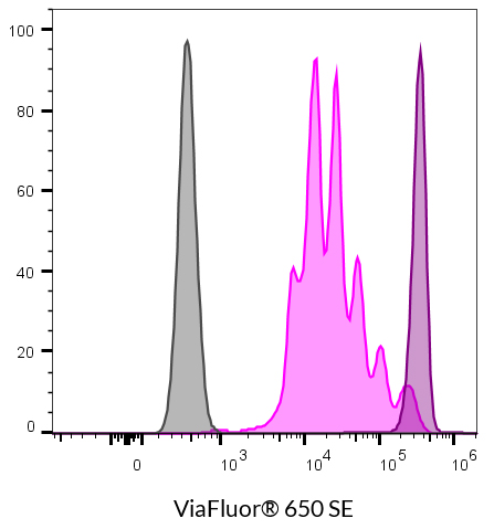 PBMCs were stained with 1 uM ViaFluor® 650 Cell Proliferation Dye, and then T cell proliferation was stimulated with CD3/CD28 T Cell Activator Beads and IL-2. After 4 days, the cells were analyzed by flow cytometry in the R660 (APC) channel. The stimulated T cell proliferation can be measured by the number of individual peaks (light pink). Unstimulated cells are shown in dark pink, and unstained cells and in gray.
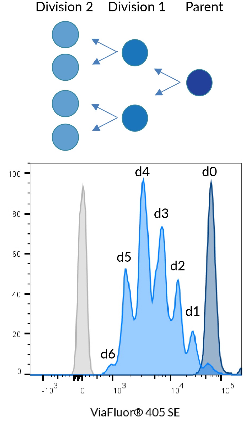 Principle of cell division tracking with ViaFluor® Cell Proliferation Dyes. When a stained cell divides, each daughter cell receives half the dye in the parent cell, with each cell division represented as a successively dimmer population on a flow cytometry histogram. Data shown using 5 uM ViaFluor® 405 to stain PBMCs.
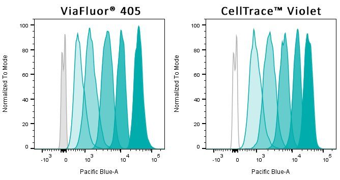 Figure 3. Jurkat cells were stained with 5 uM cell proliferation dye: ViaFluor® 405 (left panel) or CellTrace™ Violet (right panel). Cells were analyzed in the Pacific Blue channel immediately after staining, and then every day for four days. Both dyes give sharp peaks showing the progressive decrease in signal caused by cell division.
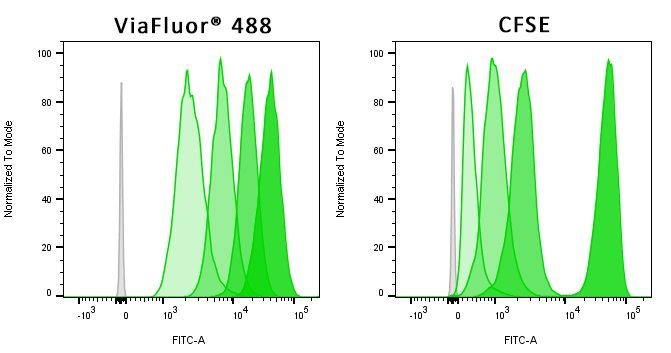 Figure 4. Jurkat cells were stained with 3 uM cell proliferation dye: ViaFluor® 488 (left panel) or ViaFluor® CFSE (right panel). Cells were analyzed in the FITC channel immediately after staining, and then every day for three days. CFSE loses a lot of signal between initial staining and Day 1, limiting the number of cell divisions that can be measured, while ViaFluor® 488 retains dye better and gives nice sharp peaks for each division cycle.
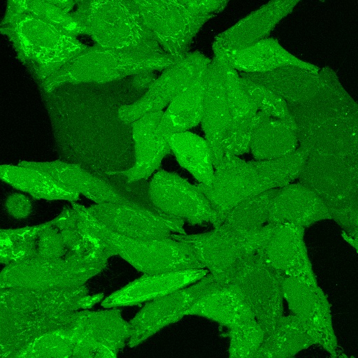 HeLa cells were cultured for 2 days, then labeled at room temperature with ViaFluor® 488 SE.
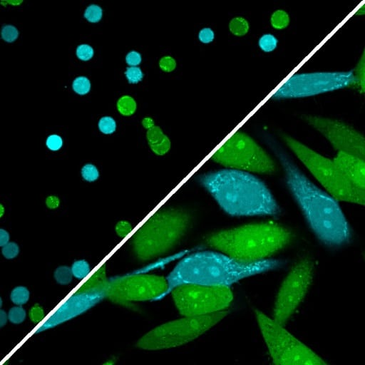 HeLa cells were labeled with either ViaFluor® 405 SE (blue) or ViaFluor® 488 SE (green) after trypsinization. The two samples were mixed together and seeded. Top left: immediately after seeding; bottom right: after 24 hours of culture.
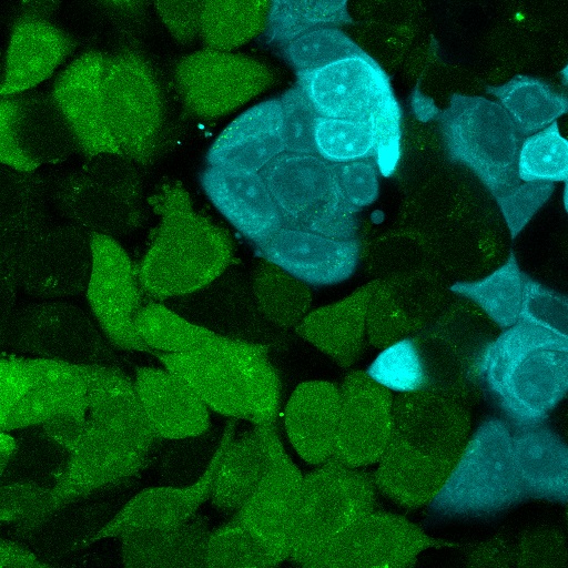 Adherent HeLa cells and MCF-7 cells were labeled at room temperature with ViaFluor® 488 (green) and ViaFluor® 405 (blue), respectively. The cells were then trypsinized, mixed, plated, and cultured for four days.
 HeLa cells were plated in a coverglass-bottom 96-well plate and cultured for 24 hours, then stained with ViaFluor® 405 SE (top row) or ViaFluor® 488 SE (bottom row) at 2 uM in HBSS for 15 minutes at 37°C. Cells were washed, placed in culture medium, and incubated at 37°C for 30 minutes, then imaged (initial staining). Labeled cells were cultured for 48 hours, then fixed with ice-cold methanol for 10 minutes at -20°C. Cells were imaged on a Zeiss LSM 700 confocal microscope using a 40X oil objective; the same laser and gain settings were used to capture images at each time point.
  Two aliquots of Jurkat cells were labeled separately with either ViaFluor® 488 SE (green) or ViaFluor® 405 SE (blue), then washed, mixed together, and imaged using an Etaluma Lumascope LS720 microscope with 40X objective.
Product Description
ViaFluor® SE Cell Proliferation Kits use amine-reactive dyes to covalently label cells throughout the cell cytoplasm and intracellular compartments for fixable fluorescent staining. Cell proliferation dyes are commonly used to monitor cell division by flow cytometry. The dyes also can be used to stably label cells to image cell morphology, or to track cell populations in mixed co-culture experiments.
Features
- Non-toxic dyes covalently label cell cytoplasm for fixable staining
- Track cell proliferation in vivo or in vitro by dye dilution using flow cytometry
- Long term imaging of cell morphology or co-cultures by microscopy
- Excellent performance & lower cost compared to leading competitors
- ViaFluor® 405, 488, and 650 are much less toxic than CFSE
Kit Components
- Lyophilized dye in single use vials
- Anhydrous DMSO for dissolving dye
Spectral Properties (Ex/Em after hydrolysis)
- ViaFluor® CFSE: 495/515 nm
- ViaFluor® 488 SE: 495/524 nm
- ViaFluor® 405 SE: 387/446 nm
- ViaFluor® 650 SE: 653/682 nm
ViaFluor® SE Dyes for Cell Proliferation Tracking
ViaFluor® SE dyes are membrane-permeant compounds that are initially non-fluorescent esters, but are converted to fluorescent dyes by intracellular esterases and will covalently react with amine groups on intracellular proteins at the same time, forming fluorescent conjugates that are retained in the cell. Immediately after staining, a single bright fluorescent population will be detected by flow cytometry. With each cell division, daughter cells inherit roughly half of the fluorescent label, allowing the number of cell divisions that occur after labeling to be detected by the appearance of successively dimmer fluorescent peaks on a flow cytometry histogram compared to cells analyzed immediately after staining. Thus, cell proliferation dyes can be used to track multiple cell divisions of cells grown in culture or injected in vivo after labeling with the ViaFluor® SE dye.
The number of assays that can be performed per kit depends on the dye concentration used (see the product protocol for more information). When used at 1 uM to label 106 cells in one mL, each dye vial can be used for 90-100 labelings.
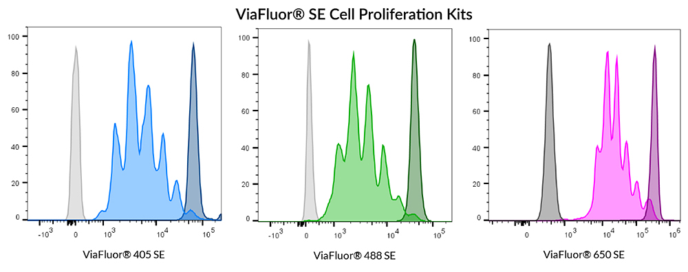
Human PBMCs were stained with ViaFluor® 405 (left), ViaFluor® 488 (center), or ViaFluor® 650 (right). Cells were stimulated with Dynabeads® Human T-Activator CD3/CD28 beads and 100 ng/mL IL-2. Cells were analyzed 4 days post-induction. CD3+ T-cells are shown. Unstimulated cells (dark peaks) and unstained cells (gray) are shown for comparison.
Other Applications for ViaFluor® SE Dyes
ViaFluor® SE dyes also can be used for imaging cell morphology or identifying cells in co-culture by microscopy. Because the staining is non-toxic and well-retained, it can be used for imaging live cells over time. See our Tech Tip: Using ViaFluor® SE Stains for Cell Tracing and Co-Culture.
All three ViaFluor® SE dyes can stain gram-positive bacteria, but not gram-negative bacteria. ViaFluor® CFSE stains the cytoplasm in yeast, but ViaFluor® 405 & ViaFluor® 488 stain the yeast cell periphery. See our Cellular Stains Table for more information on how our dyes stain various organisms.
Cell Division | Catalog No. | Ex/Em (nm) | Flow detection | Features |
|---|---|---|---|---|
| ViaFluor® 405 SE Cell Proliferation Kit | 30068 | 387/446 | Pacific Blue® | • ViaFluor® 405 SE replaces CellTrace™ Violet |
| ViaFluor® 488 SE Cell Proliferation Kit | 30086 | 495/524 | FITC | • ViaFluor® 488 SE is a unique, improved green dye to replace CFSE |
| ViaFluor® 650 SE Cell Proliferation Kit | 30139 | 653/682 | APC | • ViaFluor® 650 SE is an improved and cost-effective alternative to CellTrace™ Far Red |
| ViaFluor® CFSE Cell Proliferation Kit | 30050 | 495/519 | FITC | • Classic cell division tracing dye, see our ViaFluor® 488 SE for an improved alternative |
Biotium also offers the the ViaPlex™ 2-Color Cell Barcoding Kits for analyzing up to 15 samples in a single tube to save time and reagents, as well as Live-or-Dye™ Fixable Viability Staining Kits for robust dead cell detection in 18 colors spanning blue to near-IR. Learn more about our products for flow cytometry.
Technology

Tech Tips


 Powered by Bioz