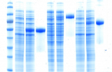
Protein staining is a fundamental technique in biochemistry and molecular biology used to visualize proteins after their separation, typically by gel electrophoresis. This process involves the application of dyes or stains that bind to proteins, producing visible bands or spots that can be analyzed both qualitatively and quantitatively. Protein staining is crucial for assessing protein expression, integrity, and relative abundance across a variety of experimental workflows.
Applications and Importance
- Protein Quantification and Normalization: Total protein stains offer a robust alternative to single housekeeping proteins, which may exhibit variable expression under certain experimental conditions. Staining the entire proteome allows for normalization based on total protein content, improving reproducibility and quantitative accuracy in western blotting and other comparative techniques.
- Protein Identification and Analysis: Following SDS-PAGE or 2D electrophoresis, protein staining enables the identification of individual protein bands. Researchers can estimate molecular weight by comparing to a protein ladder, assess sample purity, or excise specific bands for mass spectrometry-based proteomics. This is especially valuable in differential expression studies, biomarker discovery, and quality control of purified proteins.
- Detection of Post-Translational Modifications: Specialized stains can selectively bind to specific post-translational modifications (PTMs), such as phosphorylation, glycosylation, or ubiquitination. These targeted stains facilitate the study of protein function, signal transduction pathways, and disease-related modifications in proteomic and functional assays.
Protein staining continues to be an essential tool in molecular biology, proteomics, and clinical diagnostics. Whether for verifying protein presence, comparing expression levels, or preparing samples for downstream analysis, the choice of stain significantly impacts data quality and interpretability.
