Early Embryonic Development
Early embryonic development is the highly coordinated biological process through which a single fertilized cell gives rise to a multicellular embryo with distinct cell lineages and developmental potential. This period establishes the molecular and cellular foundation for all subsequent embryogenesis and is therefore central to developmental biology, reproductive medicine, regenerative medicine, and embryonic stem cell research. Because many developmental events are conserved across mammals while retaining important species-specific characteristics, discoveries from model organisms have significantly advanced our understanding of human embryo development and improved assisted reproductive technologies, including IVF embryo culture.
Early Embryonic Development Stages
Development begins with fertilization, when the sperm and oocyte fuse to form the zygote, a single totipotent cell capable of generating both embryonic and extraembryonic tissues required for complete organismal development. During the earliest stages, the embryo remains enclosed within the zona pellucida, a specialized glycoprotein matrix that protects the developing embryo and prevents premature implantation.
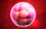
The zygote undergoes a series of rapid mitotic cleavage divisions, producing progressively smaller blastomeres without increasing the overall embryo volume. Following successive cleavage events, the embryo reaches the morula stage, typically consisting of approximately 16–32 cells. A defining feature of this stage is compaction, during which blastomeres increase cell–cell adhesion, establish apical–basal polarity, and form tighter intercellular contacts, creating the structural organization required for subsequent lineage specification.
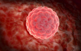
As development proceeds, fluid accumulates between the cells through a process known as cavitation, leading to the formation of the blastocoel cavity and ultimately blastocyst formation. The blastocyst represents the final stage of the preimplantation embryo and comprises two principal cellular populations: the inner cell mass (ICM), which gives rise to the embryo proper, and the surrounding trophoblast (trophectoderm), which primarily contributes to placental and other extraembryonic tissues. These sequential developmental transitions constitute the principal early embryonic development stages investigated in mammalian developmental biology and reproductive research.
Cell Fate Specification and Pluripotency
A defining characteristic of early embryonic development is lineage segregation, during which initially equivalent blastomeres progressively acquire distinct developmental identities . The first lineage decision separates the trophoblast from the ICM, while a subsequent segregation within the ICM produces the epiblast and the primitive endoderm. The epiblast ultimately forms the fetus, whereas the primitive endoderm contributes to extraembryonic tissues that support embryonic growth and development.
Cellular developmental potential changes throughout these stages. The zygote and earliest blastomeres are totipotent, whereas cells of the epiblast become pluripotent, retaining the capacity to generate all embryonic cell types while lacking the ability to produce the complete extraembryonic tissues necessary for full organismal development. These pluripotent cells provide the biological foundation for pluripotent stem cells, making the preimplantation embryo a cornerstone of embryonic stem cell research, regenerative medicine, and developmental biology.
Another universal developmental landmark is zygotic genome activation (ZGA), during which developmental control shifts from maternally deposited transcripts and proteins to transcription initiated by the embryonic genome. Although the timing of zygotic genome activation differs among mammalian species, this transition is essential for continued embryonic development and represents one of the defining molecular events of the preimplantation period.
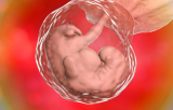
Following blastocyst expansion, the embryo hatches from the zona pellucida before implantation, when the trophoblast establishes interactions with the maternal endometrium. Implantation marks the transition from preimplantation to post-implantation development and precedes the onset of gastrulation, during which the three primary germ layers—ectoderm, mesoderm, and endoderm—begin to form, establishing the body plan for subsequent organogenesis .
Research Tools for Early Embryonic Development Studies
Advances in human embryo development, preimplantation embryo research, and stem cell biology depend on robust experimental systems and standardized developmental biology reagents that ensure reproducibility across laboratories. Widely used embryo culture media, including KSOM and sequential media designed for IVF embryo culture, provide optimized conditions for mammalian embryo development in vitro. These media are commonly supplemented with extracellular matrix coatings, growth factors such as LIF and FGF, and carefully validated small-molecule modulators that support embryo and pluripotent stem cell maintenance.
Immunophenotypic characterization remains fundamental for identifying key markers in preimplantation embryos. Frequently used antibodies include anti-OCT4, anti-SOX2, and anti-NANOG for pluripotency; anti-SSEA-3, anti-SSEA-4, anti-TRA-1-60, and anti-TRA-1-81 for human pluripotent stem cell characterization; and anti-E-cadherin for evaluating compaction and epithelial organization . Researchers also routinely employ pluripotent stem cell identification kits, immunofluorescence staining kits, embryo viability and quality assessment assays, CRISPR-based genome editing tools, and advanced embryo imaging reagents for high-resolution live-cell analysis.
Together, these specialized antibodies, culture systems, molecular reagents, and analytical kits provide the experimental foundation for investigating mammalian embryogenesis with high precision and reproducibility. As imaging, genome editing, and stem cell technologies continue to evolve, these integrated research tools will further advance our understanding of early embryonic development and accelerate discoveries in developmental biology, reproductive medicine, and regenerative medicine.