


Glutamine synthetase antibody



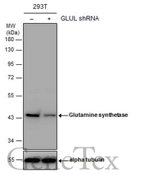
Non-transfected (–) and transfected (+) 293T whole cell extracts (30 μg) were separated by 10% SDS-PAGE, and the membrane was blotted with Glutamine synthetase antibody (GTX109121) diluted at 1:1000. The HRP-conjugated anti-rabbit IgG antibody (GTX213110-01) was used to detect the primary antibody.
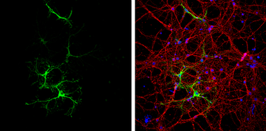
Glutamine synthetase antibody detects Glutamine synthetase protein at astrocytes by immunofluorescent analysis.
Sample: DIV9 rat E18 primary cortical neurons were fixed in 4% paraformaldehyde at RT for 15 min.
Green: Glutamine synthetase protein stained by Glutamine synthetase antibody (GTX109121) diluted at 1:500.
Red: beta Tubulin 3/ Tuj1, a neuron cell marker, stained by beta Tubulin 3/ Tuj1 antibody [GT11710] (GTX631836) diluted at 1:500.
Blue: Fluoroshield with DAPI (GTX30920).
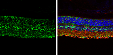
Glutamine synthetase antibody detects Glutamine synthetase protein expression by immunohistochemical analysis.
Sample:Paraffin-embedded adult mouse retina.
Green: Glutamine synthetase protein stained by Glutamine synthetase antibody (GTX109121) diluted at 1:250.
Red: beta Tubulin 3/ TUJ1, stained by beta Tubulin 3/ TUJ1 antibody [GT11710] (GTX631836) diluted at 1:250.
Blue: Fluoroshield with DAPI (GTX30920).
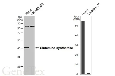
Various whole cell extracts (30 μg) were separated by 10% SDS-PAGE, and the membrane was blotted with Glutamine synthetase antibody (GTX109121) diluted at 1:1000. The HRP-conjugated anti-rabbit IgG antibody (GTX213110-01) was used to detect the primary antibody. Corresponding RNA expression data for the same cell lines are based on Human Protein Atlas program.
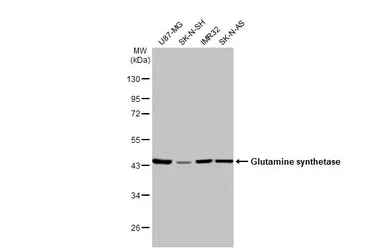
Various whole cell extracts (30 μg) were separated by 10% SDS-PAGE, and the membrane was blotted with Glutamine synthetase antibody (GTX109121) diluted at 1:1000. The HRP-conjugated anti-rabbit IgG antibody (GTX213110-01) was used to detect the primary antibody.
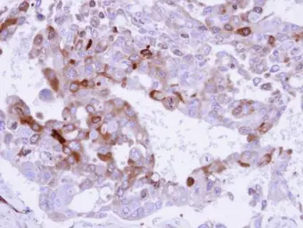
Immunohistochemical analysis of paraffin-embedded H441 xenograft , using Glutamine Synthetase (GTX109121) antibody at 1:500 dilution.
Antigen Retrieval: Trilogy™ (EDTA based, pH 8.0) buffer, 15min
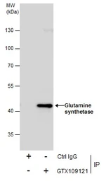
Immunoprecipitation of Glutamine synthetase protein from IMR32 whole cell extracts using 5 μg of Glutamine synthetase antibody (GTX109121).
Western blot analysis was performed using Glutamine synthetase antibody (GTX109121).
EasyBlot anti-Rabbit IgG (GTX221666-01) was used as a secondary reagent.
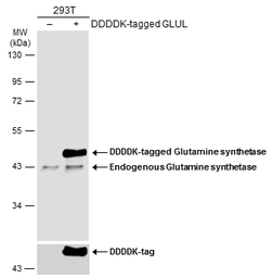
Non-transfected (–) and transfected (+) 293T whole cell extracts (30 μg) were separated by 10% SDS-PAGE, and the membrane was blotted with Glutamine synthetase antibody (GTX109121) diluted at 1:10000. The HRP-conjugated anti-rabbit IgG antibody (GTX213110-01) was used to detect the primary antibody.
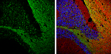
Glutamine synthetase antibody detects Glutamine synthetase protein expression by immunohistochemical analysis.
Sample: Frozen-sectioned adult mouse cerebellum.
Green: Glutamine synthetase protein stained by Glutamine synthetase antibody (GTX109121) diluted at 1:250.
Red: beta Tubulin 3/ TUJ1, stained by beta Tubulin 3/ TUJ1 antibody [GT11710] (GTX631836) diluted at 1:500.
Blue: Fluoroshield with DAPI (GTX30920).
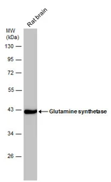
Rat tissue extract (50 μg) was separated by 10% SDS-PAGE, and the membrane was blotted with Glutamine synthetase antibody (GTX109121) diluted at 1:50000.
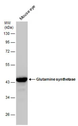
Mouse tissue extract (50 μg) was separated by 10% SDS-PAGE, and the membrane was blotted with Glutamine synthetase antibody (GTX109121) diluted at 1:20000.
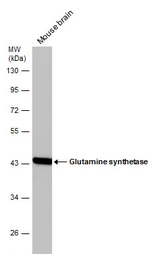
Mouse tissue extract (50 μg) was separated by 10% SDS-PAGE, and the membrane was blotted with Glutamine synthetase antibody (GTX109121) diluted at 1:50000.
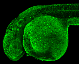
Glutamine synthetase antibody detects Glutamine synthetase protein on zebrafish by whole mount immunohistochemical analysis.
Sample: 2 days-post-fertilization zebrafish embryo.
Glutamine synthetase antibody (GTX109121) dilution: 1:100.
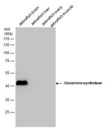
Glutamine synthetase antibody detects Glutamine synthetase protein by Western blot analysis. Zebrafish tissue extracts (30 μg) was separated by 10 % SDS-PAGE, and the membrane was blotted with Glutamine synthetase antibody (GTX109121) at a dilution of 1:10000.
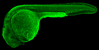
Glutamine synthetase antibody detects Glutamine synthetase protein on zebrafish by whole mount immunohistochemical analysis.
Sample: 1 day-post-fertilization zebrafish embryo.
Glutamine synthetase antibody (GTX109121) dilution: 1:100.
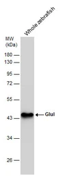
Whole zebrafish extract (30 μg) was separated by 10% SDS-PAGE, and the membrane was blotted with Glutamine synthetase antibody (GTX109121) diluted at 1:10000.
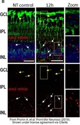
The data was published in the journal Front Mol Neurosci in 2019. PMID: 30930743
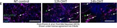
The data was published in the journal Front Mol Neurosci in 2019. PMID: 30930743
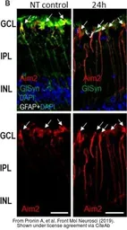
The data was published in the journal Front Mol Neurosci in 2019. PMID: 30930743
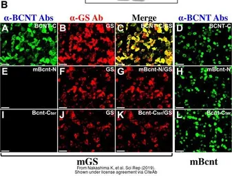
The data was published in the journal Sci Rep in 2019. PMID: 31616018
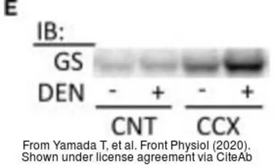
The data was published in the journal Front Physiol in 2020.PMID: 32425814
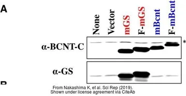
The data was published in the journal Sci Rep in 2019. PMID: 31616018
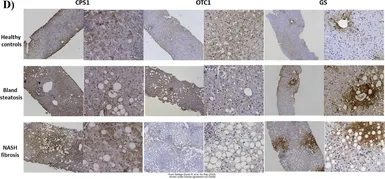
The data was published in the 2022 in Sci Rep. PMID: 35232986