

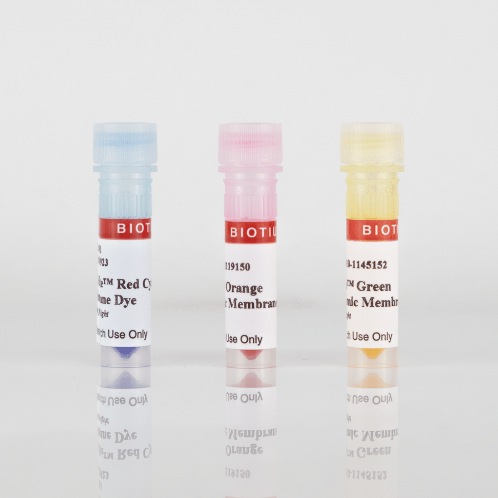
CellBrite® Red Cytoplasmic Membrane Dye

Cat# 30023
Size : 1ML
Marca : Biotium
CellBrite® Cytoplasmic Membrane Dyes
Dye solutions of lipophilic carbocyanine dyes DiB, Neuro-DiO, DiI, and DiD, as well as novel near-infrared lipophilic dyes for non-toxic labeling of cytoplasmic membranes.



Product Description
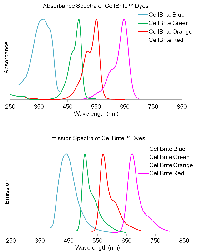
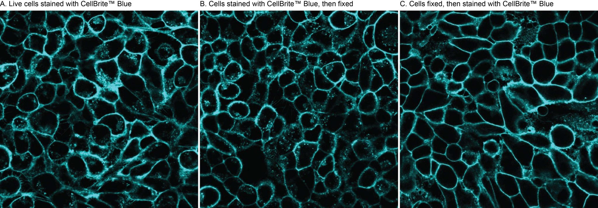
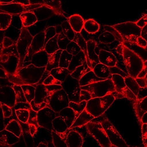
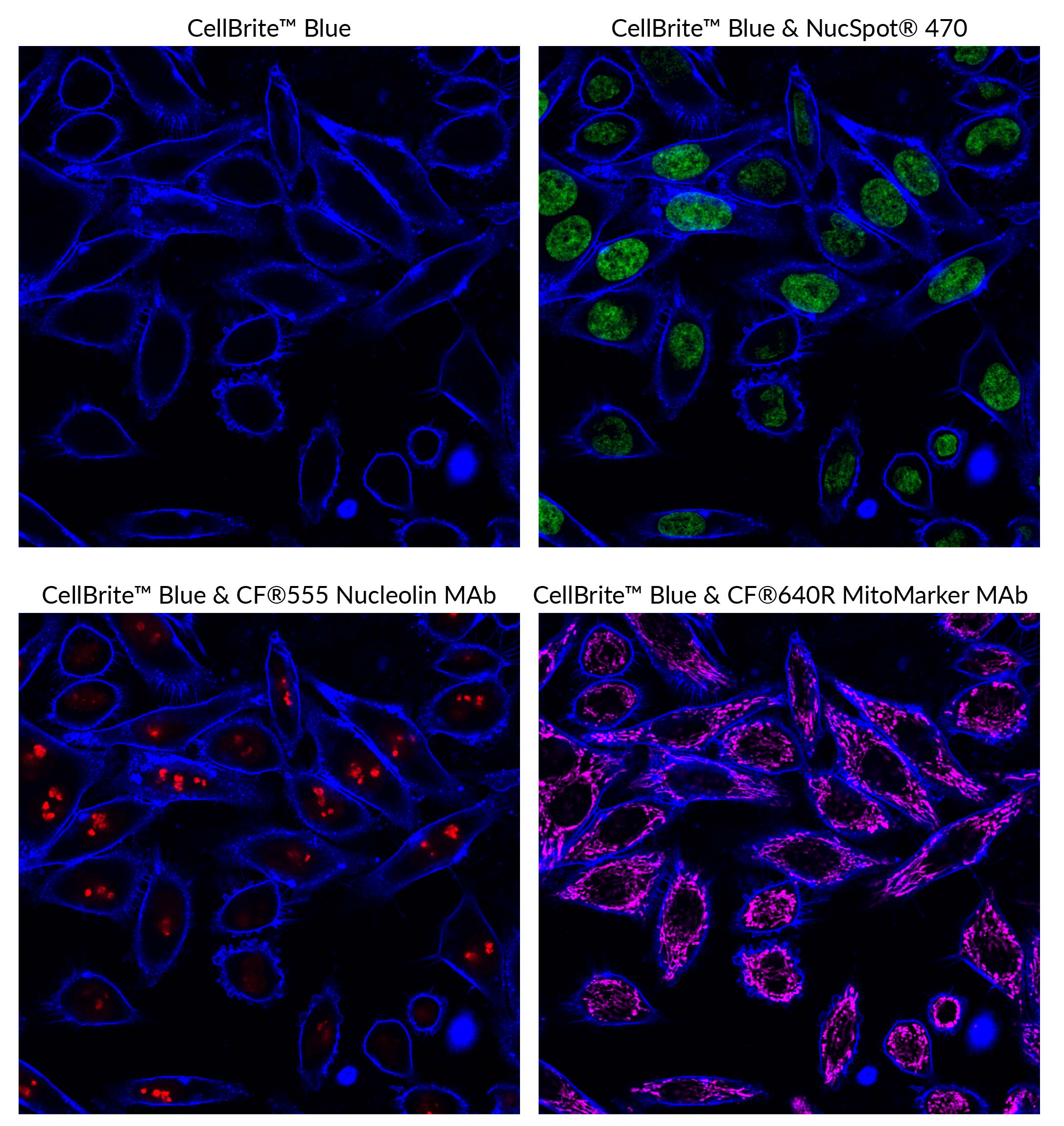
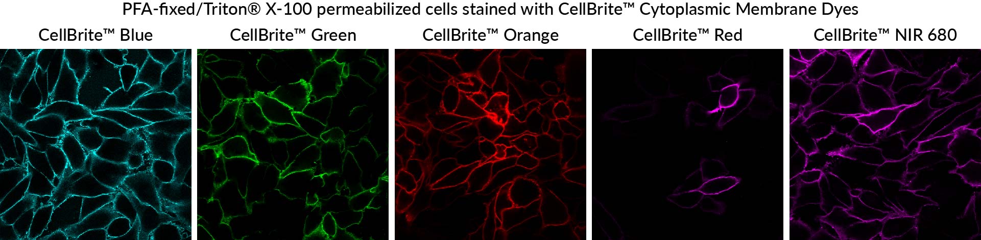
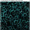
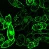
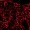
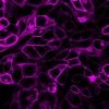
CellBrite® Cytoplasmic Membrane Labeling Kits can be used to label cell cytoplasmic membranes with fluorescent dyes, available in blue, green, orange, red, and near-infrared. The labeling is nontoxic and suitable for long-term tracking of cells. The dyes also can be used to stain membranes of formaldehyde-fixed cells.
Features
- Ready-to-use solutions of DiB, neuro-DiO, DiI, DiD and novel near-infrared lipophilic dyes for membrane staining
- Low toxicity and minimal dye transfer between cells
- Useful for cell tracing, tracking, and transplantation studies
- Formaldehyde-fixable
- Stain live cells in culture medium, or stain formaldehyde-fixed cells
Kit Components
- Optimized dye stock solution
- Loading buffer (CellBrite® Blue only)
CellBrite® Cytoplasmic Membrane Stains are available as classic lipophilic carbocyanine dyes and novel near-infrared lipophilic dyes. CellBrite® Blue is based on DiB, CellBrite® Green is based on Neuro-DiO, CellBrite® Orange is based on DiI, and CellBrite® Red is based on DiD. These carbocyanine dyes label cytoplasmic membrane and intracellular membrane structures efficiently and permanently. They have been used as tracers in cell fusion, cell adhesion, and cell migration applications due to their properties of low cytotoxicity and high resistance to intercellular transfer. By combining multiple CellBrite® Cytoplasmic Membrane Stains, one can label multiple cell populations with different colors for studies of cell-cell interactions.
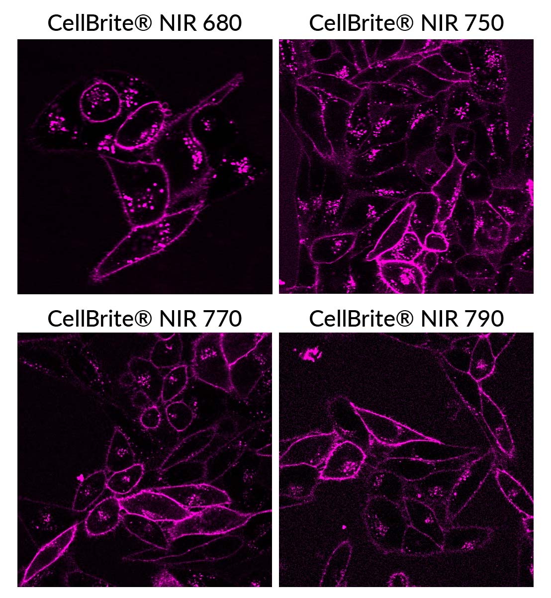
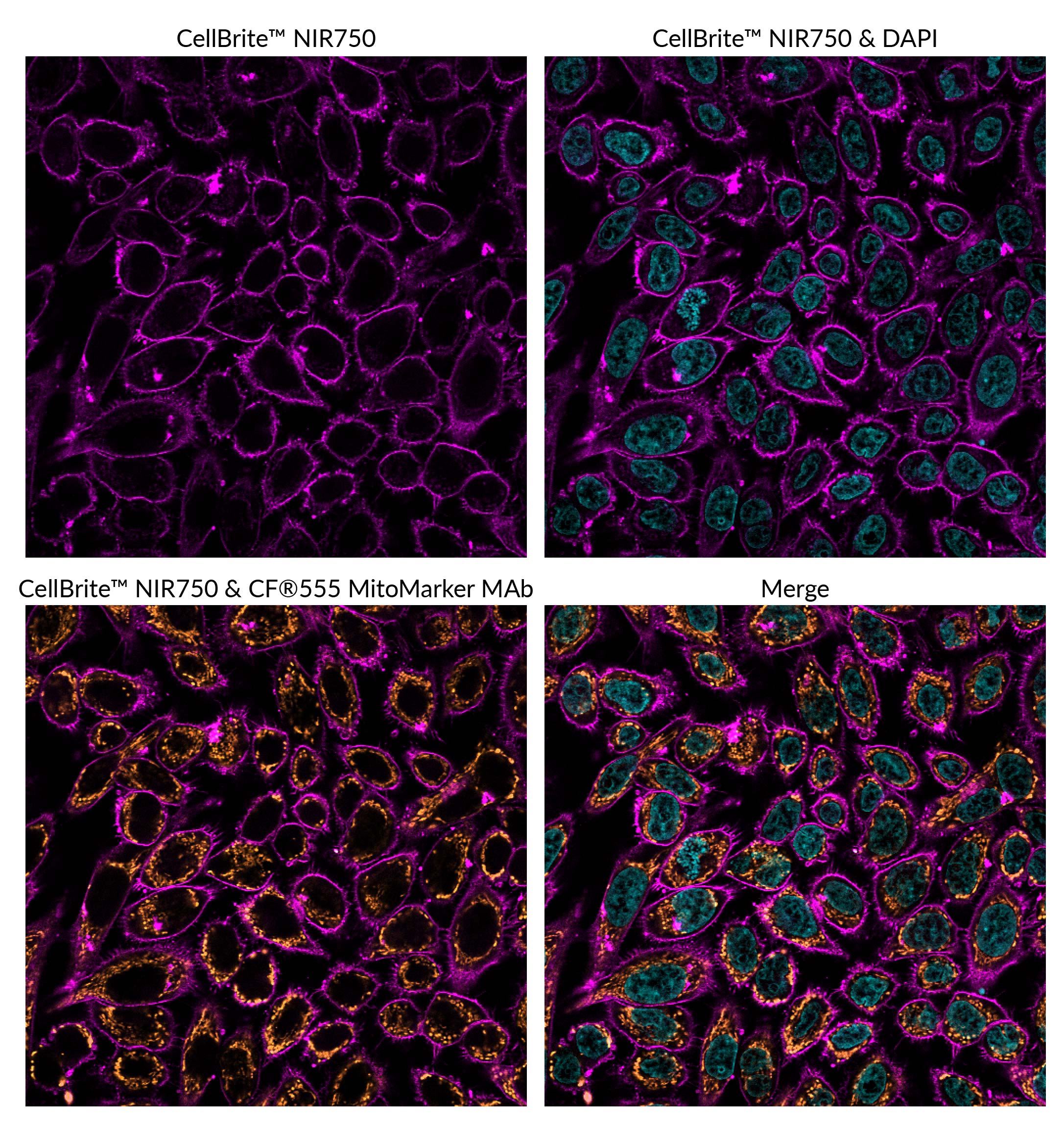
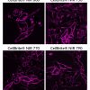
The near-infrared CellBrite® NIR Cytoplasmic Membrane Dyes are unique and due to their long wavelength emission, they can be used to label cells for small animal imaging studies for non-invasive imaging of cell migration and cell homing. CellBrite® NIR Dyes have long 18-carbon hydrophobic tails and an additional water-soluble group. These unique chemical structure elements make the dyes easy to dissolve while providing highly stable cytoplasmic membrane staining, unlike traditional carbocyanine dyes like DiI, DiO, and DiR, which are often difficult to dissolve or prone to precipitation during cell staining.
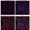
Biotium's CellBrite® Cytoplasmic Membrane Dyes are ready-to-use and can be added directly to normal culture media to uniformly label suspended or adherent cells in culture. CellBrite® Dyes allow cell populations to be marked in distinctive fluorescent colors for identification after mixing. Double labeling can identify cells that have fused or formed stable clusters.
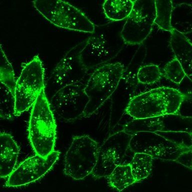
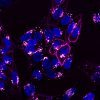
Note: CellBrite® Dyes primarily localize to cell surface membranes shortly after staining. However, in live cells the stained membranes become internalized by endocytosis, so staining becomes primarily intracellular over the course of a few hours in culture. See "Find the Right Stain for Your Application" below, and our Membrane Staining & Imaging Tech Tip to learn more.
Find the Right Stain for Your Application
CellBrite® Dyes are non-toxic and stain cells very stably. They can be used to track cells for days to weeks. However, over time the dyes will be internalized by endocytosis, resulting in labeling of intracellular vesicles. A few hours after staining, the dyes will no longer outline the plasma membrane, but will be localized inside the cell. For long-term imaging of cell surfaces in culture we recommend our CellBrite® Steady Membrane Staining Kits which distribute between the cell surface and intracellular compartments, so cells retain cell surface staining over time. For long-term visualization of cell morphology in culture, our stable, non-toxic ViaFluor® SE Cell Proliferation Kits may also be a suitable alternative. ViaFluor® SE dyes covalently label cells throughout the cytoplasm and can be tracked for several days or longer by microscopy or flow cytometry. See our Tech Tip: Using ViaFluor® SE Stains for Cell Tracing and Co-Culture to learn more.
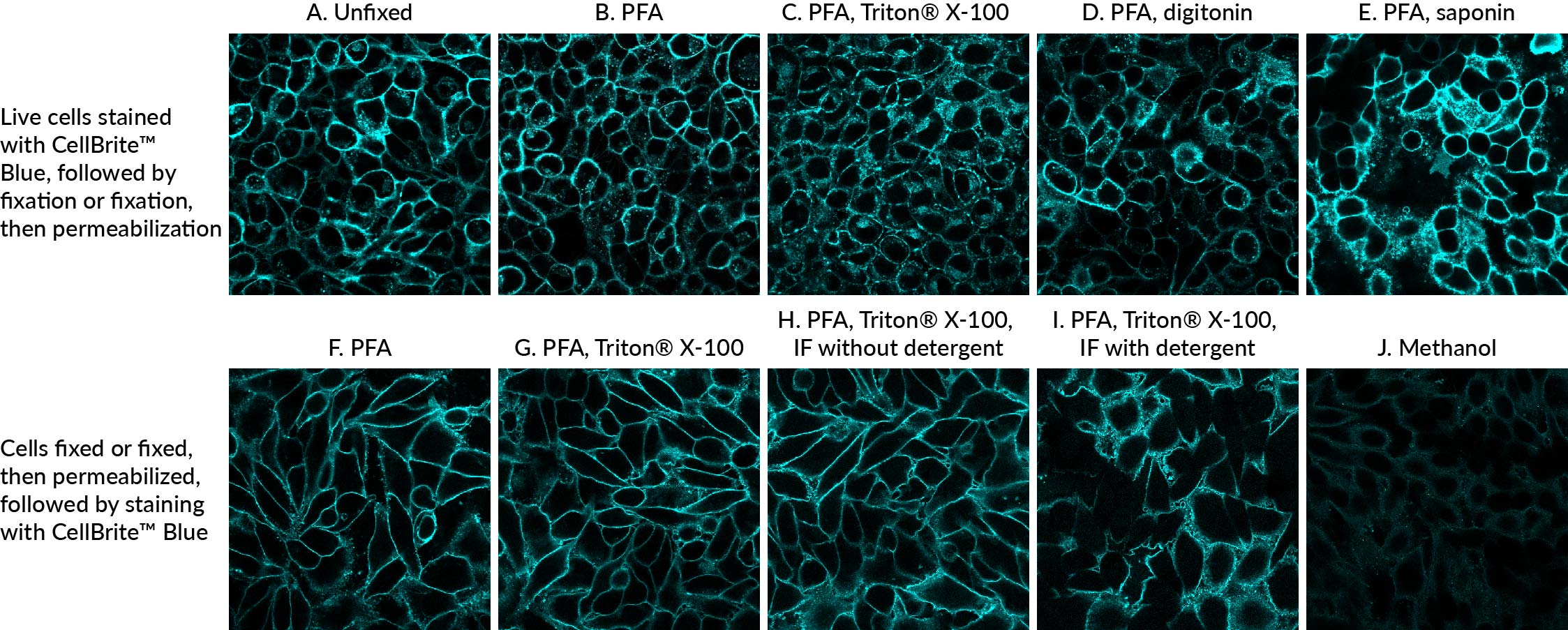
CellBrite® Dyes can be used to stain formaldehyde-fixed cells, but the staining has poor tolerance for methanol fixation or detergent permeabilization. However, permeabilizing fixed cells before staining with CellBrite® Dyes can give good results, see our Tech Tip: Combining Lipophilic Membrane Dyes with Immunofluorescence. For staining formaldehyde-fixed cells we strongly recommend using CytoLiner™ Fixed Cell Membrane Stains which offer more robust and consistent results over the CellBrite® Cytoplasmic Membrane Dyes.
For membrane stains that withstand fixation with either formaldehyde or methanol, as well as detergent permeabilization after staining, see our GlycoLiner™ Cell Surface Glycoprotein Labeling Kits, CellBrite® Fix Membrane Stains and MemBrite® Fix Cell Surface Staining Kits.
CellBrite® Cytoplasmic Membrane Dyes do not stain yeast or bacteria. See our Cellular Stains Table for more information on how our dyes stain various organisms.
Watch our video where Technical Applications Scientist II, Jacqueline Steenhuis PhD answers your top questions about Biotium's various membrane stains for fluorescence microscopy.
For additional support or product recommendations, contact us at techsupport@biotium.com.
CellBrite® Cytoplasmic Membrane Dyes
| Dye | Ex/Em | Format | Size | Catalog No. |
|---|---|---|---|---|
| CellBrite® Blue | 360/441 nm | 250 uL of Labeling Solution, 200 X in DMSO 250 uL of Cell Loading Buffer, 200X in DMSO | 50 assays | 30024 |
| CellBrite® Green | 489/506 nm | 200X in EtOH | 1 mL | 30021 |
| CellBrite® Orange | 551/569 nm | 200X in EtOH | 1 mL | 30022 |
| CellBrite® Red | 648/670 nm | 200X in EtOH | 1 mL | 30023 |
| CellBrite® NIR680 | 683/724 nm | 2 mM in DMSO* | 100 uL | 30070 |
| CellBrite® NIR750 | 748/781 nm | 2 mM in DMSO* | 100 uL | 30077 |
| CellBrite® NIR770 | 767/806 nm | 2 mM in DMSO* | 100 uL | 30078 |
| CellBrite® NIR790 | 787/820 nm | 2 mM in DMSO* | 100 uL | 30079 |
Technology

Tech Tips

Product Attributes
Probe cellular localization
For live or fixed cells
Assay type/options
Fixation options
Colors
Documents, Protocols, SDS and COA

COA
Request the Certificate of AnalysisReferences
Download a list of curated CellBrite® and MemBrite® references.
Citations
Download a list of curated CellBrite® and MemBrite® references.
 Powered by Bioz
© 2026
Powered by Bioz
© 2026