

ECL Chemiluminescent Substrate Detection Kit (Hypersensitive)

Cat# K1231-500
Size : 500mL(250mLeachforAandB)
Marca : APExBIO Technology
ECL Chemiluminescent Substrate Detection Kit (Hypersensitive)
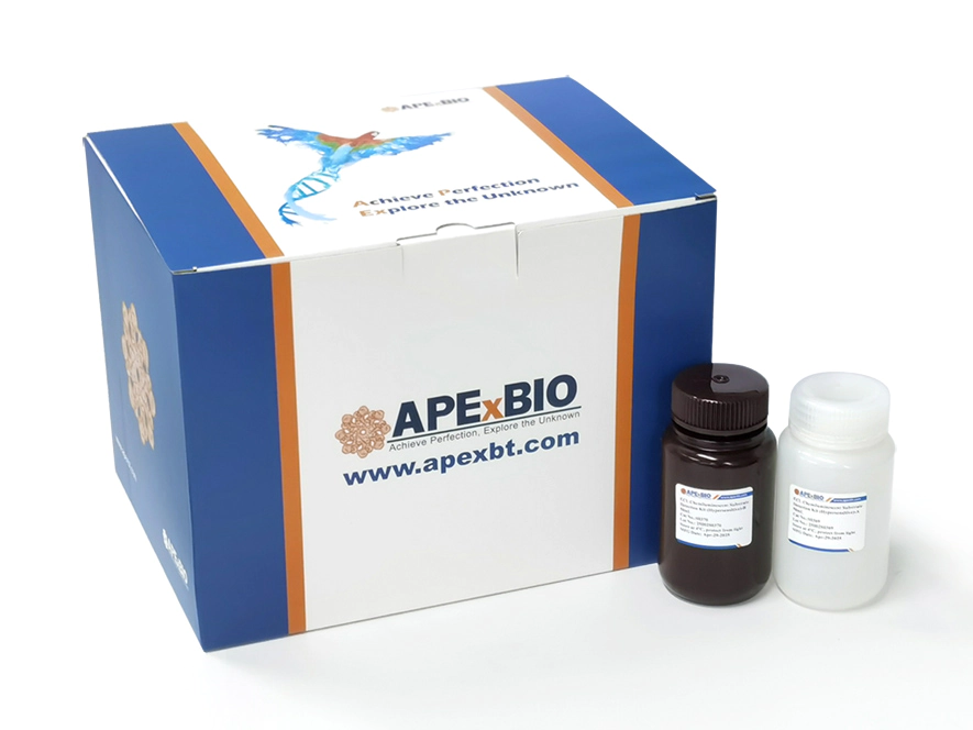
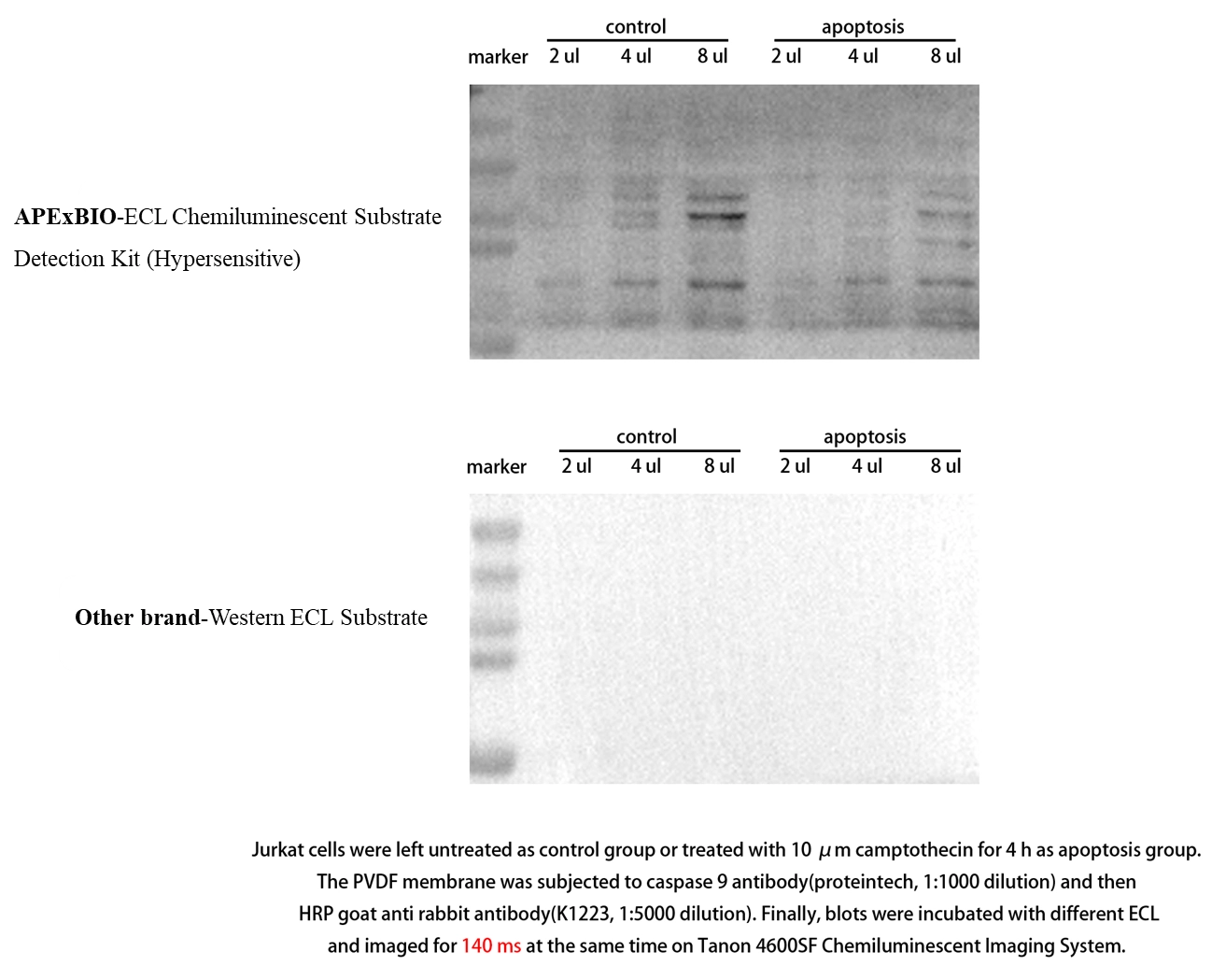
ECL Chemiluminescent Substrate Detection Kit (Hypersensitive), using HRP-mediated oxidation to generate chemiluminescent signals, is for immunoblotting to detect antigens on nitrocellulose/PVDF membranes, with high sensitivity (low picogram range) for low-abundance proteins. Its signals persist for 6-8 hours under optimal conditions, and the mixed working reagent is stable for 24 hours. It has an extended room-temperature shelf-life, and compared to conventional kits (e.g., from T and M), it shows lower background and longer signal duration.
Product Features
(1) Enhanced chemiluminescent substrate for horseradish peroxidase (HRP).
(2) Low picogram sensitivity - Detects low picogram protein bands on nitrocellulose or PVDF membranes.
(3) Long signal duration - Under optimized conditions, substrate-incubated blots can consistently output a detectable light signal for 6 to 8 hours.
(4) Stable reagent - The working solution remains stable for 24 hours; The kit can be stored at room temperature for up to 1 year.
(5) Cost-effective - Optimized for diluted antibody concentrations.
- 1. Ruoshui Li, Gaoyang Li, et al. "Targeting the ENO1 metabolic checkpoint via microenvironment-responsive nanotherapeutics promotes postinfarction cardiac repair." Sci Adv. 2026 Oct 2;12(40):eaeg3270. PMID: 42826207
- 2. Li Zhu, Shuo Chen, et al. "AFF4 Loss Compromises Osteogenesis of Orofacial Mesenchymal Stem Cells During Alveolar Bone Repair via AMPK‐Mediated Mitophagy." Cell Prolif. 2026 Sep 29:e70284. PMID: 42811695
- 3. Yaqin Tian, Liting Lu, et al. "The Hcp2b protein of APEC induces defective autophagy in HD11 cells by causing lysosomal dysfunction." Avian Pathol. 2026 Sep 11:1-14. PMID: 42725459
- 4. Meng Li, Jiayu Wang, et al. "The epigenetic regulator ALKBH5 facilitates the early adaptation of hepatocellular carcinoma cells to sorafenib treatment." J Chemother. 2026 Sep 3:1-17. PMID: 42691501
- 5. Chenfei Zhao, Xiao Liu, et al. "Amplification-free Detection of N6-Methyladenosine (m6A) Based on Droplet Digital CRISPR-Cas13a." Talanta. 2026 Jul 26;312(Pt A):130370. PMID: 42520632
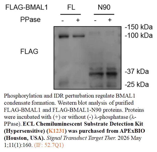
- 6. Wenzhen Gao, Li Zhu, et al. "BMAL1 regulates circadian rhythms via phase separation–mediated transcriptional hub formation." Signal Transduct Target Ther. 2026 May 1;11(1):160. PMID: 42062246
Related Biological Data
Related Biological Data
Related Biological Data
Related Biological Data
| Components | 100 mL | 500 mL |
| ECL Chemiluminescent Substrate Detection Kit (Hypersensitive)-A | 50 mL | 250 mL |
| ECL Chemiluminescent Substrate Detection Kit (Hypersensitive)-B | 50 mL | 250 mL |
Store the components dry at 4 °C and protect from light for 12 months. | ||
1. What causes an inverted image on the film, i.e., black background with white bands?
A: This is usually caused by an excessive amount of HRP in the system, which rapidly depletes the substrate. It is recommended to dilute the HRP-conjugated secondary antibody at least 10-fold to effectively resolve this issue.
2. What causes brown or yellow bands on the membrane?
A: This phenomenon is often related to suboptimal chemiluminescent development conditions, such as overly long substrate incubation or exposure times, or substrate degradation/precipitation under light. Shortening the development/exposure time and optimizing the detection conditions can alleviate this issue.
3. If the chemiluminescent signal lasts less than 8 hours, what could be the reason?
A: An excessively short signal duration is usually caused by too much HRP in the system, which rapidly depletes the substrate and leads to quick signal decay. The solution is to dilute the HRP-conjugated secondary antibody at least 10-fold.
4. If the signal is weak or absent, what are the possible causes and how can this be resolved?
A: Common causes and corresponding solutions include:
· Excessive HRP in the system (substrate depleted quickly, signal decays rapidly): dilute the HRP-conjugated secondary antibody at least 10-fold.
· Insufficient amount of antigen or antibody: increase the concentration of antigen or antibody accordingly.
· Low protein transfer efficiency: optimize transfer conditions (e.g., transfer time, current/voltage, buffer composition).
· Reduced HRP or substrate activity: perform activity testing to confirm and replace inactivated reagents.
5. If the background is too high, what are the possible causes and how can this be resolved?
A: Common causes of high background and corresponding solutions include:
· Excessive HRP in the system: dilute the HRP-conjugated secondary antibody at least 10-fold.
· Insufficient blocking: optimize blocking conditions, such as extending blocking time, adjusting blocking temperature, or shaking speed.
· Inappropriate blocking reagent: try different blocking reagents, such as non-fat dry milk, BSA, or serum.
· Inadequate washing: increase the number of washes, extend washing time, or increase the volume of wash buffer.
· Overexposure of the film: shorten the exposure time and, if necessary, use a background-reduction reagent.
· Excessive antigen or antibody concentration: reduce the amount of antigen or antibody accordingly.
6. What causes spots within the protein bands, and how can this be resolved?
A: Common causes and corresponding solutions include:
· Uneven or inefficient protein transfer: optimize the transfer procedure, e.g., check transfer voltage, time, and buffer composition to ensure uniform transfer.
· Uneven membrane hydration: fully equilibrate/hydrate the membrane according to the manufacturer's instructions to avoid local dryness or insufficient hydration.
· Air bubbles between the film and membrane: carefully remove any bubbles before exposure to ensure close contact between the membrane and film.
7. What causes speckled background on the film, and how can this be resolved?
A: Speckled background is usually caused by aggregates present in the HRP-conjugated secondary antibody solution. The solution is to filter the secondary antibody working solution through a 0.2 µm filter before use to remove aggregates.
8. What causes non-specific bands, and how can this be resolved?
A: Common causes and corresponding solutions include:
· Excessive HRP in the system: dilute the HRP-conjugated secondary antibody at least 10-fold to reduce non-specific binding signals.
· SDS-induced non-specific protein binding: avoid using SDS during the detection steps (antibody incubation and chemiluminescent detection).
9. How can the activity of the HRP/substrate system be tested?
A: The activity can be tested as follows:
· In a darkroom, prepare 1–2 mL of substrate working solution in a clean test tube.
· Turn off the lights.
· Add 1 µL of undiluted HRP-conjugated secondary antibody working solution.
Expected result: If the system is active, the solution should immediately emit blue light, and the blue signal will gradually fade over the next few minutes.