

ECL Western Blotting Substrate

Cat# MBS9633871-100mL
Size : 100mL
Marca : MyBiosource
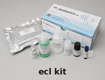
ECL Kit
For Research Use Only. Not for use in diagnostic procedures. Catalog #: MBS9633871The Affinity ECL Western Blotting Substrate MBS9633871 is a highly sensitive nonradioactive, enhanced luminol-based chemiluminescent substrate for the detection of horseradish peroxidase (HRP) on immunoblots. MBS9633871 enables the detection of picogram or… More details
Product Overview
Product Name
ECL, Kit
Full Product Name
ECL Western Blotting Substrate
Product Gene Name
ECL kit
Preparation and Storage
Upon receipt store at room temperature
Supply Chain Verification
Manufactured in a lab with traceable raw materials manufactured on site. Coordinated product portfolio of antibodies, pairs, conjugates, recombinant proteins, and immunoassay materials available, please inquire.
Info
•ECL Substrate requires more dilute antibody concentrations than those used with precipitating colorimetric HRP substrates. To optimize antibody concentrations, perform a systematic dot blot analysis.
•Empirical testing is essential to determine the appropriate blocking reagent for each Western blot system, as crossreactivity of the blocking reagent with antibodies causes nonspecific signal. Blocking buffer also affects system sensitivity.
•Avoid using milk as a blocking reagent when using avidin/biotin systems because milk contains variable amounts of endogenous biotin, which causes high background signal.
•Use sufficient volumes of wash buffer, blocking buffer, antibody solution and substrate working solution to cover blot and ensure that it never becomes dry. Using large blocking and wash buffer volumes minimizes nonspecific signal.
•For optimal results, use a shaking platform during incubation steps.
•Add Tween-20 Detergent (final concentration of 0.05-0.1%) to the blocking buffer and all diluted antibody solutions to minimize nonspecific signal.
•Do not use sodium azide as a preservative for buffers. Sodium azide is an inhibitor of HRP and could interfere with this system.
•Do not handle membrane with bare hands. Always wear gloves or use clean forceps.
•All equipment must be clean and free of foreign material. Metallic devices (e.g., scissors) must have no visible signs of rust. Rust may cause speckling and high background.
•Exposure to the sun or any other intense light can harm the substrate. For best results keep the substrate working solution in an amber bottle and avoid prolonged exposure to any intense light. Short-term exposure to typical laboratory lighting will not harm the working solution.
•Empirical testing is essential to determine the appropriate blocking reagent for each Western blot system, as crossreactivity of the blocking reagent with antibodies causes nonspecific signal. Blocking buffer also affects system sensitivity.
•Avoid using milk as a blocking reagent when using avidin/biotin systems because milk contains variable amounts of endogenous biotin, which causes high background signal.
•Use sufficient volumes of wash buffer, blocking buffer, antibody solution and substrate working solution to cover blot and ensure that it never becomes dry. Using large blocking and wash buffer volumes minimizes nonspecific signal.
•For optimal results, use a shaking platform during incubation steps.
•Add Tween-20 Detergent (final concentration of 0.05-0.1%) to the blocking buffer and all diluted antibody solutions to minimize nonspecific signal.
•Do not use sodium azide as a preservative for buffers. Sodium azide is an inhibitor of HRP and could interfere with this system.
•Do not handle membrane with bare hands. Always wear gloves or use clean forceps.
•All equipment must be clean and free of foreign material. Metallic devices (e.g., scissors) must have no visible signs of rust. Rust may cause speckling and high background.
•Exposure to the sun or any other intense light can harm the substrate. For best results keep the substrate working solution in an amber bottle and avoid prolonged exposure to any intense light. Short-term exposure to typical laboratory lighting will not harm the working solution.
Procedure Summary
Note: Antigen and antibody amounts may require optimization.
1. Dilute the primary antibody to 5.0-0.5ug/mL.
2. Dilute the secondary antibody to 1.0-0.067ug/mL.
3. Mix Solution II and I at a 1:1 ratio and add it to the blot. Incubate blot for 1-600 seconds. Note that Solution II includes Picogram and Femtogram. Please choose one according to the experimental results. It is recommended to select Solution II(picogram) firstly. If you want to enhance the effect, you can try Solution II(femtogram).
4. Drain excess reagent. Cover blot with a clear plastic sheet protector or clear plastic wrap.
5. Expose blot to X-ray film.
1. Dilute the primary antibody to 5.0-0.5ug/mL.
2. Dilute the secondary antibody to 1.0-0.067ug/mL.
3. Mix Solution II and I at a 1:1 ratio and add it to the blot. Incubate blot for 1-600 seconds. Note that Solution II includes Picogram and Femtogram. Please choose one according to the experimental results. It is recommended to select Solution II(picogram) firstly. If you want to enhance the effect, you can try Solution II(femtogram).
4. Drain excess reagent. Cover blot with a clear plastic sheet protector or clear plastic wrap.
5. Expose blot to X-ray film.
Product Categories/Family for ECL kit
ISO Certification
Manufactured in an ISO 9001:2015 Certified Laboratory.
Applications Tested/Suitable for ECL kit
Western Blot (WB)
Application Notes for ECL kit
Western Blotting Procedure
1. Remove blot from the transfer apparatus and block nonspecific sites with Blocking Reagent for 60 minutes at room temperature (RT) with shaking. If desired, block overnight at 2-8°C without shaking.
2. Remove the Blocking Reagent and add the primary antibody working dilution. Incubate blot for 1 hour at RT with shaking or overnight at 2-8°C without shaking.
3. Briefly rinse membrane in Wash Buffer two times.
4. Wash membrane by suspending it in Wash Buffer and agitating for > 5 minutes. Replace Wash Buffer at least 4-6 times. Increasing the Wash Buffer volume, the number of washes and wash duration may help minimize background signal.
5. Incubate blot with the HRP-conjugate working dilution for 1 hour at RT with shaking.
6. Repeat Steps 3 and 4 to remove nonbound HRP-conjugate.
Note: Membrane MUST be thoroughly washed after incubation with the HRP-conjugate.
7. Prepare the substrate working solution by mixing equal parts of Detection Reagents 1 and 2. Use 0.125mL Working Solution per cm2 of membrane.
Note: For best results prepare working solution immediately before use. The working solution is stable for 1 hour at RT.
8. Incubate blot with working solution for 1 minute at RT.
9. Remove blot from working solution and place it in a plastic sheet protector or clear plastic wrap. Use an absorbent tissue to remove excess liquid and to carefully press out any bubbles from between the blot and the membrane protector.
10. Place the protected membrane in a film cassette with the protein side facing up. Turn off all lights except those appropriate for X-ray film exposure (e.g., a red safelight).
Note: Film must remain dry during exposure. For optimal results, perform the following precautions:
• Make sure excess substrate is removed from the membrane and the membrane protector.
• Use gloves during the entire film-handling process.
• Never place a blot on developed film, as chemicals on the film may reduce signal.
11. Carefully place X-ray film on top of the membrane. Perform a first-time exposure of 30 seconds. Vary the exposure time to achieve optimal results.
12. Develop film using appropriate developing solution and fixative. If signal is too intense, reduce exposure time or strip and re-probe the blot with decreased antibody concentrations.
1. Remove blot from the transfer apparatus and block nonspecific sites with Blocking Reagent for 60 minutes at room temperature (RT) with shaking. If desired, block overnight at 2-8°C without shaking.
2. Remove the Blocking Reagent and add the primary antibody working dilution. Incubate blot for 1 hour at RT with shaking or overnight at 2-8°C without shaking.
3. Briefly rinse membrane in Wash Buffer two times.
4. Wash membrane by suspending it in Wash Buffer and agitating for > 5 minutes. Replace Wash Buffer at least 4-6 times. Increasing the Wash Buffer volume, the number of washes and wash duration may help minimize background signal.
5. Incubate blot with the HRP-conjugate working dilution for 1 hour at RT with shaking.
6. Repeat Steps 3 and 4 to remove nonbound HRP-conjugate.
Note: Membrane MUST be thoroughly washed after incubation with the HRP-conjugate.
7. Prepare the substrate working solution by mixing equal parts of Detection Reagents 1 and 2. Use 0.125mL Working Solution per cm2 of membrane.
Note: For best results prepare working solution immediately before use. The working solution is stable for 1 hour at RT.
8. Incubate blot with working solution for 1 minute at RT.
9. Remove blot from working solution and place it in a plastic sheet protector or clear plastic wrap. Use an absorbent tissue to remove excess liquid and to carefully press out any bubbles from between the blot and the membrane protector.
10. Place the protected membrane in a film cassette with the protein side facing up. Turn off all lights except those appropriate for X-ray film exposure (e.g., a red safelight).
Note: Film must remain dry during exposure. For optimal results, perform the following precautions:
• Make sure excess substrate is removed from the membrane and the membrane protector.
• Use gloves during the entire film-handling process.
• Never place a blot on developed film, as chemicals on the film may reduce signal.
11. Carefully place X-ray film on top of the membrane. Perform a first-time exposure of 30 seconds. Vary the exposure time to achieve optimal results.
12. Develop film using appropriate developing solution and fixative. If signal is too intense, reduce exposure time or strip and re-probe the blot with decreased antibody concentrations.
Other Notes
Small volumes of ECL kit vial(s) may occasionally become entrapped in the seal of the product vial during shipment and storage. If necessary, briefly centrifuge the vial on a tabletop centrifuge to dislodge any liquid in the container`s cap. Certain products may require to ship with dry ice and additional dry ice fee may apply.
Searchable Terms for ECL purchase
MBS9633871 is a kit targeting ECL. Also known as ECL Western Blotting Substrate.
Additional Details
NCBI/Uniprot data below describe general gene information for ECL. It may not necessarily be applicable to this product.
NCBI Accession #
Protein Family
Eclosion hormone
Precautions
All of MyBioSource's Products are for scientific laboratory research purposes and are not for diagnostic, therapeutics, prophylactic or in vivo use. Through your purchase, you expressly represent and warrant to MyBioSource that you will properly test and use any Products purchased from MyBioSource in accordance with industry standards. MyBioSource and its authorized distributors reserve the right to refuse to process any order where we reasonably believe that the intended use will fall outside of our acceptable guidelines.
Disclaimer
While every efforts were made to ensure the accuracy of the information provided in this datasheet, MyBioSource will not be liable for any omissions or errors contained herein. MyBioSource reserves the right to make changes to this datasheet at any time without prior notice.
It is the responsibility of the customer to report product performance issues to MyBioSource within 30 days of receipt of the product. Please visit our Terms & Conditions page for more information.
It is the responsibility of the customer to report product performance issues to MyBioSource within 30 days of receipt of the product. Please visit our Terms & Conditions page for more information.
Product Note
Select online data sheet information is drawn from bioinformatics databases, occasionally resulting in ambiguous or non-relevant product information. It is the responsibility of the customer to review, verify, and evaluate the information to make sure it matches their requirements before purchasing the kit. Our ELISA Kit assays are dynamic research tools and sometimes they may be updated and improved. If the format of this assay is important to you then please request the current manual or contact our technical support team with a presales inquiry before placing an order. We will confirm the current details of the assay. We cannot guarantee the sample manual posted online is the most current manual, it is intended to serve as an example only. Please refer to the instructions For Use provided with the assay kit for precise details.
Si può anche essere interessati nei seguenti prodotti:
Cat#
Descrizione
Cond.
Priced