


The RayBiotech Lipid Peroxidation (MDA) Assay Kit provides a simple, reproducible, and standardized tool for assaying lipid peroxidation in serum, plasma, cell or tissue lysates, urine, or other body fluid samples.
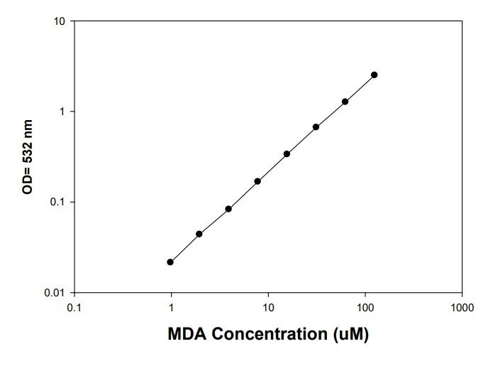
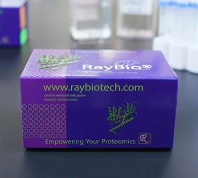
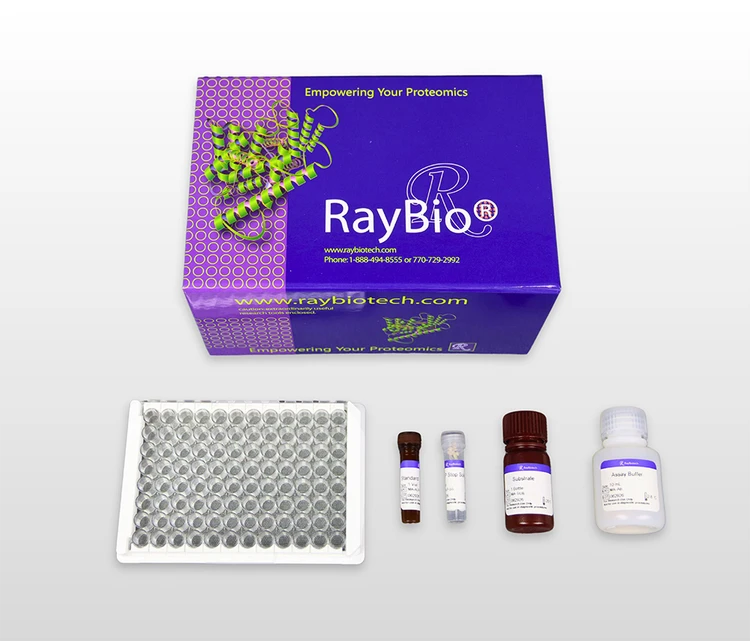
Standard curve graph of lipid peroxidation assay measuring MDA concentration.
Lipid Peroxidation (MDA) Assay Kit
Contact Us for Bulk Request
Product Description
Specifications
| Size | 2 Plate Kit, 4 Plate Kit |
| Sensitivity | 0.38 µM |
| Compatible Sample Types | Plasma, Serum, Tissue Lysates, Cell Lysates, Urine |
| Quantitative/Semi-Quantitative | Quantitative |
| Solid Support | 96-well Microplate |
| Method Of Detection | Colorimetric |
| Research Area | Cardiovascular Disease, Inflammation, Oxidative Stress, Ferroptosis, Lipid Peroxidation |
| Shipping Type | Blue ice |
| Storage | 2-8°C |
| Storage / Stability | The entire kit may be stored at 2-8 °C for up to 6 months from the date of shipment. For prepared reagent storage, see manual. |
Related Products
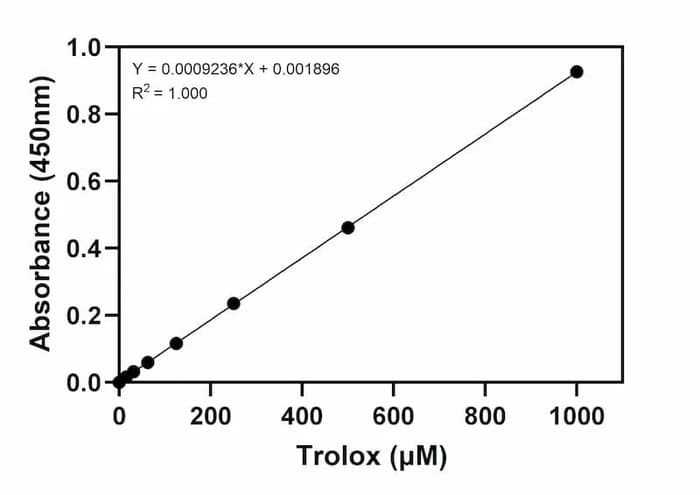
Total Antioxidant Capacity (TAC) Assay Kit (Colorimetric)
As low as $299.00
Total Thiol Quantification Assay Kit
As low as $299.00
Introduction
Oxidative stress can lead to toxic effects by generating peroxides and free radicals that can damage all cell components, including proteins, lipids, and DNA. Lipid peroxidation is a well-known mechanism of cellular damage in both animals and plants and has been associated with cardiovascular diseases, atherosclerosis, diabetes, liver disorders, and inflammatory rheumatic diseases. Thiobarbituric Acid Reactive Substances (TBARS) are byproducts of lipid peroxidation which are caused by reactive oxygen species and other oxidative mechanisms. Malondialdehyde (MDA) is the major TBARS and is widely used as an indicator of oxidative stress in cells and tissues.
The RayBio Lipid Peroxidation (MDA) Assay Kit provides a simple, reproducible, and standardized tool for assaying lipid peroxidation in serum, plasma, cell or tissue lysates, urine, or other body fluid samples. The assay utilizes the reaction of MDA with thiobarbituric acid (TBA), producing a pink-colored product. The intensity of the pink color, measured at 532 nm, is directly proportional to the concentration of MDA in the sample. The MDA standard or unknown samples containing MDA are first reacted with TBA under high temperature (95°C) and acidic conditions. The MDA-TBA adduct is then measured colorimetrically at 532 nm. The MDA content in unknown samples is determined by comparison with the MDA standard curve.
Kit Components
| Component | Size / Description |
|---|---|
| Microplates | Two 96-well plates (12 strips x 8 wells) |
| MDA Standard | 1 vial (25 µl) |
| Thiobarbituric Acid (TBA) | 1 bottle (1g white powder) |
| SDS Solution | 20 ml |
| 2X TBA Acid Diluent | 25 ml |
| Sodium Hydroxide Solution | 5 ml |
| 100X BHT Solution | 1 vial (1 ml) |
Additional Materials Required
- 1.5 ml microcentrifuge tubes and 50 ml conical tubes
- Benchtop centrifuge and microcentrifuge
- Water bath or heat block capable of 95°C
- Distilled or deionized water
- Precision pipettes to deliver 2 µl to 1 ml volumes
- Adjustable 1-25 ml pipettes for reagent preparation
- Spectrophotometric microplate reader capable of reading at 532 nm
- Analytical balance
Assay Procedure Summary
Prepare and mix all reagents thoroughly before use. Each MDA-containing sample and standard should be assayed in duplicate. High content MDA samples can be further diluted for analysis.
- Pipette 100 µl of samples or prepared MDA series standards (see Pre-Assay Preparation, B) to each microcentrifuge tube.
- Add 100 µl of the SDS Solution to each sample and the MDA standards. Mix thoroughly and quick spin, Incubate samples for 5 minutes at room temperature.
- Add 250 µl of prepared TBA Reagent (see Pre-Assay Preparation, A-3) to each sample and standard. Mix thoroughly then incubate at 95°C for 60 minutes.
- Put all tubes on ice and incubate for 5 minutes.
- Centrifuge all tubes at 1600 x g for 10 minutes.
- Transfer 200 µL of supernatant from each sample and the MDA standards to a 96 well microplate. Read the absorbance at 532 nm immediately.
citations
Evaluation of Oxidative Stress Markers in Post-Surgical Head and Neck Cancer Patients Rehabilitated with Removable Prosthetic Restorations
- Year:
- 2025
- Journal Name:
- Antioxidants
- PubMed ID:
- 41300442
Haematococcus pluvialis extract YHC-T-2414 mitigates exercise- induced fatigue by promoting antioxidant defense and mitochondrial biogenesis
- Species:
- Mouse
- Sample Type:
- Serum (ICR mice, exercise fatigue model)
- Year:
- 2026
- Journal Name:
- Frontiers in Nutrition
- PubMed ID:
- 42656621
Exercise decreases stress induced bladder hyperalgesia, voiding frequency and oxidative load
- Sample Type:
- Tissue Lysate / Homogenate (bladder lysates); Urine
- Year:
- 2026
- Journal Name:
- Scientific Reports
Multiomics analysis unveils an inosine-sensitive DNA damage response in neurogenic bladder after spinal cord injury
- Species:
- Rat
- Sample Type:
- Tissue Lysate / Homogenate (bladder mesenchymal cells)
- Year:
- 2025
- Journal Name:
- JCI Insight
- PubMed ID:
- 40338665
COPZ1 depletion induces cell death through different mechanisms in in vitro murine models of thyroid cancer
- Species:
- Mouse
- Sample Type:
- Cell Lysate (T4888M, 3868 cell lysates); Tissue Lysate / Homogenate
- Year:
- 2026
- Journal Name:
- European thyroid journal
- PubMed ID:
- 42605848
Transcriptional Control by FOXO4 Regulates Trophoblast Differentiation and Safeguards Placental Development Through MITD1- and PCBP2-Mediated Redox Homeostasis
- Year:
- 2026
- Journal Name:
- FASEB Journal
- PubMed ID:
- 42201761
Influence of ApoE genotype on doxorubicin-induced cognitive impairment in juvenile rats
- Species:
- Rat
- Sample Type:
- Tissue Lysate / Homogenate (cortex lysates)
- Year:
- 2025
- Journal Name:
- Scientific Reports
- PubMed ID:
- 41436539
Insight into Nephrotoxicity and Processing Mechanism of Arisaema erubescens (Wall.) Schott by Metabolomics and Network Analysis
- Species:
- Rat
- Sample Type:
- kidney
- Year:
- 2023
- Journal Name:
- Drug Design Development and Therapy
SARS-CoV-2 Accessory Protein Orf7b Induces Lung Injury via c-Myc Mediated Apoptosis and Ferroptosis
- Species:
- Human
- Sample Type:
- Cell Lysate (Orf7b-overexpressing lung epithelial cells); Tissue Lysate / Homogenate
- Year:
- 2024
- Journal Name:
- International Journal of Molecular Sciences
- PubMed ID:
- 38256231
Melatonin attenuates radiation-induced cortical bone-derived stem cells injury and enhances bone repair in postradiation femoral defect model
- Species:
- Mouse
- Sample Type:
- Other / Unspecified (cortical bone-derived stem cells CBSCs)
- Year:
- 2021
- Journal Name:
- Military Medical Research
- PubMed ID:
- 34895335