

LipidSpot™ 610 Lipid Droplet Stain

Cat# 70069
Size : 125UL
Marca : Biotium
LipidSpot™ Lipid Droplet Stains
LipidSpot™ dyes rapidly stain lipid droplets in live cells or fixed cells, with no wash step required. Available with green or red/far-red fluorescence.
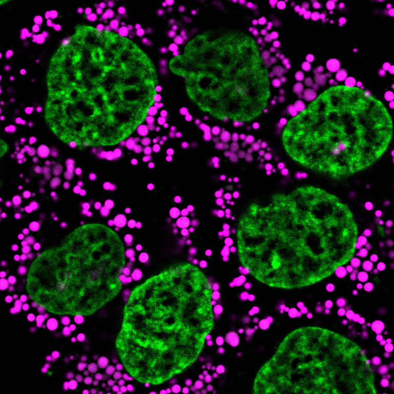 Formaldehyde-fixed, permeabilized HeLa cells stained with LipidSpot™ 610 Lipid Droplet Stain (magenta) and NucSpot® 470 Nuclear Stain (green).
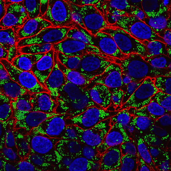 Figure 6. Fixed oleic-acid treated HeLa cells stained with LipidSpot™ 488 (lipid droplets, green) CF®594 WGA (cell surface, red), and Hoechst 33258 (nuclei, blue).
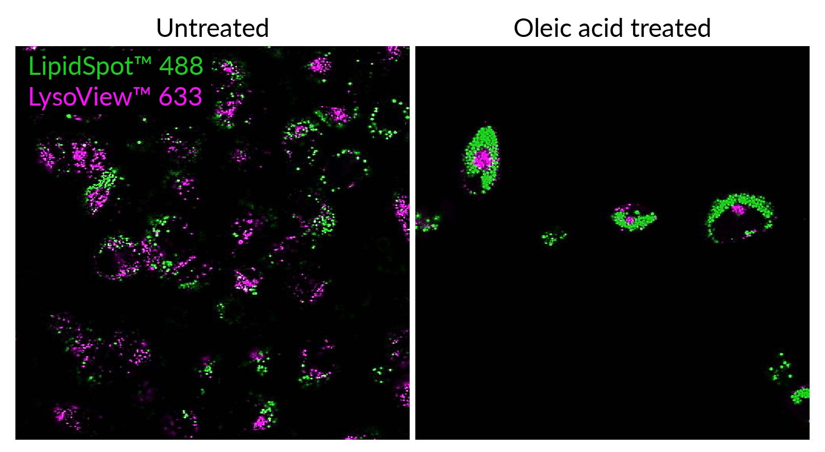 Untreated and oleic acid-treated HeLa cells co-stained with LipidSpot™ 488 (green) and LysoView™ 633 lysosome stain (magenta), showing distinct localization of the two stains.
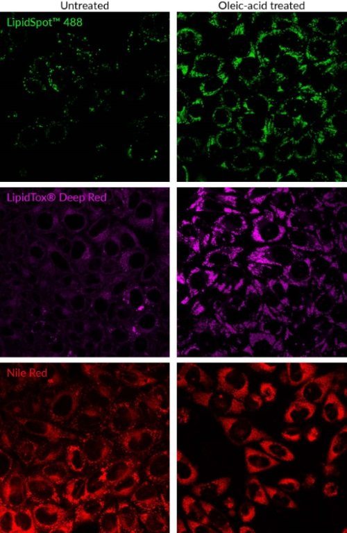 Comparison of lipid droplet dyes. HeLa cells were left untreated or treated with oleic acid to induce lipid droplet formation. Cells were stained live with LipidSpot 488 or Nile Red, or fixed and stained with LipidTox Deep Red (Thermo Fisher).
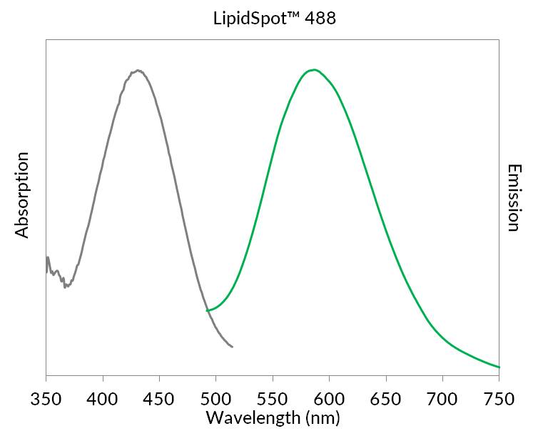 Absorption and emission spectra of LipidSpot™ 488 dye in vegetable oil. The spectral properties of the dye in cells are similar.
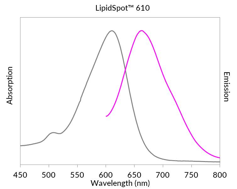 Absorption and emission spectra of LipidSpot™ 610 dye in vegetable oil. In cells, the excitation and emission spectra are green-shifted (left-shifted) 20-25 nm.
Product Description
LipidSpot™ stains are fluorescent dyes that rapidly stain lipid droplets in live or fixed cells, with no wash step and minimal background.
- Rapidly and specifically stain lipid droplets
- Minimal background, no wash required
- Stain live or fixed cells, or fix and permeabilize after staining
- Suitable for staining 3D cell spheroids
- Available with green or red/far-red fluorescence
- LipidSpot™ 488 validated in super-resolution imaging by SIM
- Supplied at 1000X in DMSO
Intracellular lipid droplets are cytoplasmic organelles involved in the storage and regulation of triglycerides and cholesterol esters. LipidSpot™ dyes are fluorogenic neutral lipid stains that rapidly accumulate in lipid droplets, where they become brightly fluorescent. The dyes can be used to stain lipid droplets in both live and fixed cells, with no wash step required. Cells also can be fixed and permeabilized after staining. LipidSpot™ stains show minimal background staining of cellular membranes or other organelles, unlike traditional dyes like Nile Red.
Find the Right Stain for Your Application
LipidSpot™ 488 has excitation around 430 nm, and can be excited equally well at 405 nm or 488 nm. In cells, it stains lipid droplets with bright green fluorescence detectable in the FITC channel. LipidSpot™ 488 has been validated in super-resolution imaging by SIM (Ref. 3), and for staining of 3-D cell spheroids (Ref. 7).
LipidSpot™ 610 has excitation/emission at ~592/638 nm in cells; it is optimally detected in the Texas Red® channel, but is also bright in the Cy®3 and far-red Cy®5 channels. Therefore, we don’t recommend pairing LipidSpot™ 610 with other red or far-red probes.
In yeast, LipidSpot™ 488 stains intracellular membranes, but LipidSpot™ 610 does not. In bacteria, both LipidSpot™ dyes can stain gram-positive but not gram-negative strains. See our Cellular Stains Table for more information on how our dyes stain various organisms.
LipidSpot™ Lipid Droplet Stains
| LipidSpot™ Stain | Abs/Em | Detection channel | Catalog no. | Size (1000X in DMSO) |
|---|---|---|---|---|
| LipidSpot™ 488 | 427/585 nm (in vegetable oil or cells) | FITC, GFP | 70065-T | 20 uL |
| 70065 | 125 uL | |||
| LipidSpot™ 610 | 610/663 nm (in vegetable oil) ~592/638 nm (in cells) | Texas Red® or Cy®5 | 70069-T | 20 uL |
| 70069 | 125 uL |
Technology

Highlighted Citations

Tech Tips


 Powered by Bioz