

NucSpot® Live 488 Nuclear Stain, Trial Size

Cat# 40081-T
Size : 1KIT
Marca : Biotium
NucSpot® Live Cell Nuclear Stains
No-wash, fluorescent DNA nuclear stains with low toxicity for long-term live cell imaging or fixed cell staining. Choose green or far-red fluorescence.
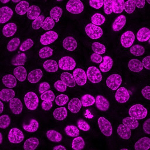
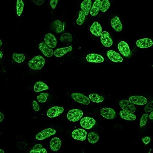
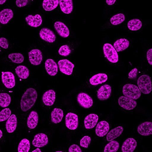
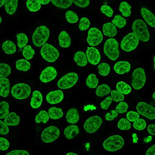 Formaldehyde-fixed, Triton X-100-permeabilized HeLa cells stained with NucSpot® Live 488.
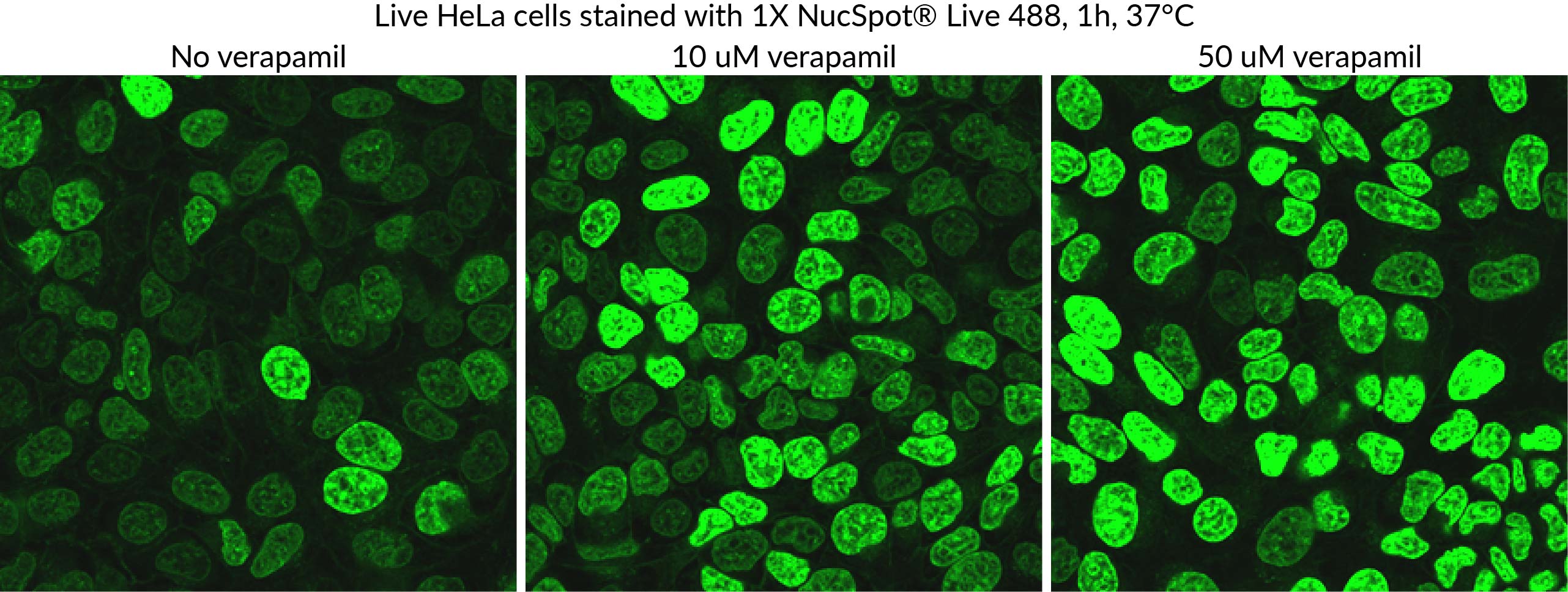 Comparison of NucSpot® Live 488 staining of live HeLa cells with or without verapamil. Verapamil can enhance dye retention in some cell types, leading to brighter and more even staining.
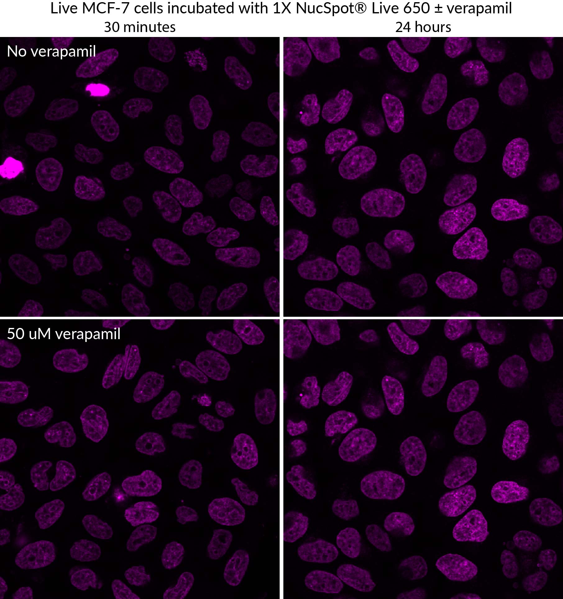 Comparison of NucSpot® Live 650 staining of live MCF-7 cells with or without verapamil. Dye and verapamil were added at time 0, and incubated with the cells continuously for 24 hours at 37°C. In some cell types, verapamil can enhance dye retention, leading to brighter and more even staining.
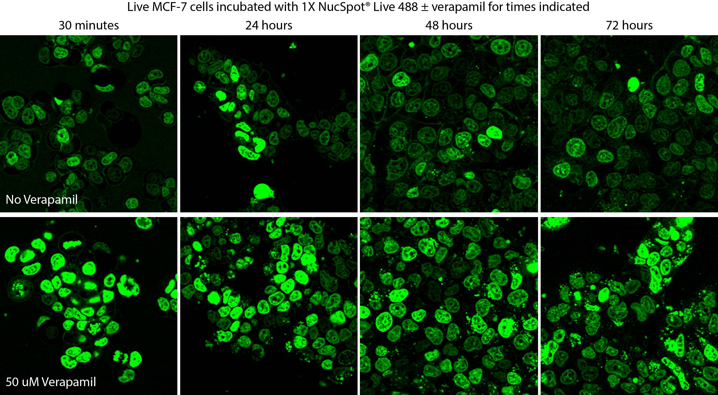 Time course of live MCF-7 cell staining with NucSpot® Live 488, with or without verapamil. Dye and verapamil were added at time 0 and incubated with cells continuously for three days. Verapamil improves dye retention and staining. NucSpot® Live 488 with verapamil showed no obvious toxicity in MCF-7 cells over the course of 72 hours. With longer incubation times, some extranuclear staining can be observed.
 Time-lapse movie of wild-type nuclei stained with NucSpot® Live 650 as they deform against rhodamine-labeled microposts (red) (Scale bar = 5 µm). Credit: Katiyar et al. https://doi.org/10.1002/advs.202201248 reproduced under the Creative Commons license.
 3D tissue dynamics of branching mouse mammary gland organoids. Organoids were incubated with NucSpot® Live 650 and treated with growth factors to trigger post-natal remodeling. The organoids were imaged for 48 hours on the Leica Viventis SCAPE light sheet microscope. Credit: Ayushi Bhanushali, and Sergio P. Acebrón.
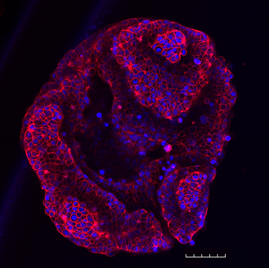 T47-D spheroid stained with NucSpot® Live 650 (nuclei, blue) and CellBrite® Steady 594 (membranes, red). T47-D cells were plated at 5,000 cells per well in a LICORbio 3D Essentials Kit ULA 96-well plate and cultured for 48 hours. The spheroid was stained with dyes in complete medium for 30 minutes without a wash step and imaged on an Evident FV4000 confocal microscope with 20X objective to create a tile-stitched 3D projection image; scale bar: 100 um.
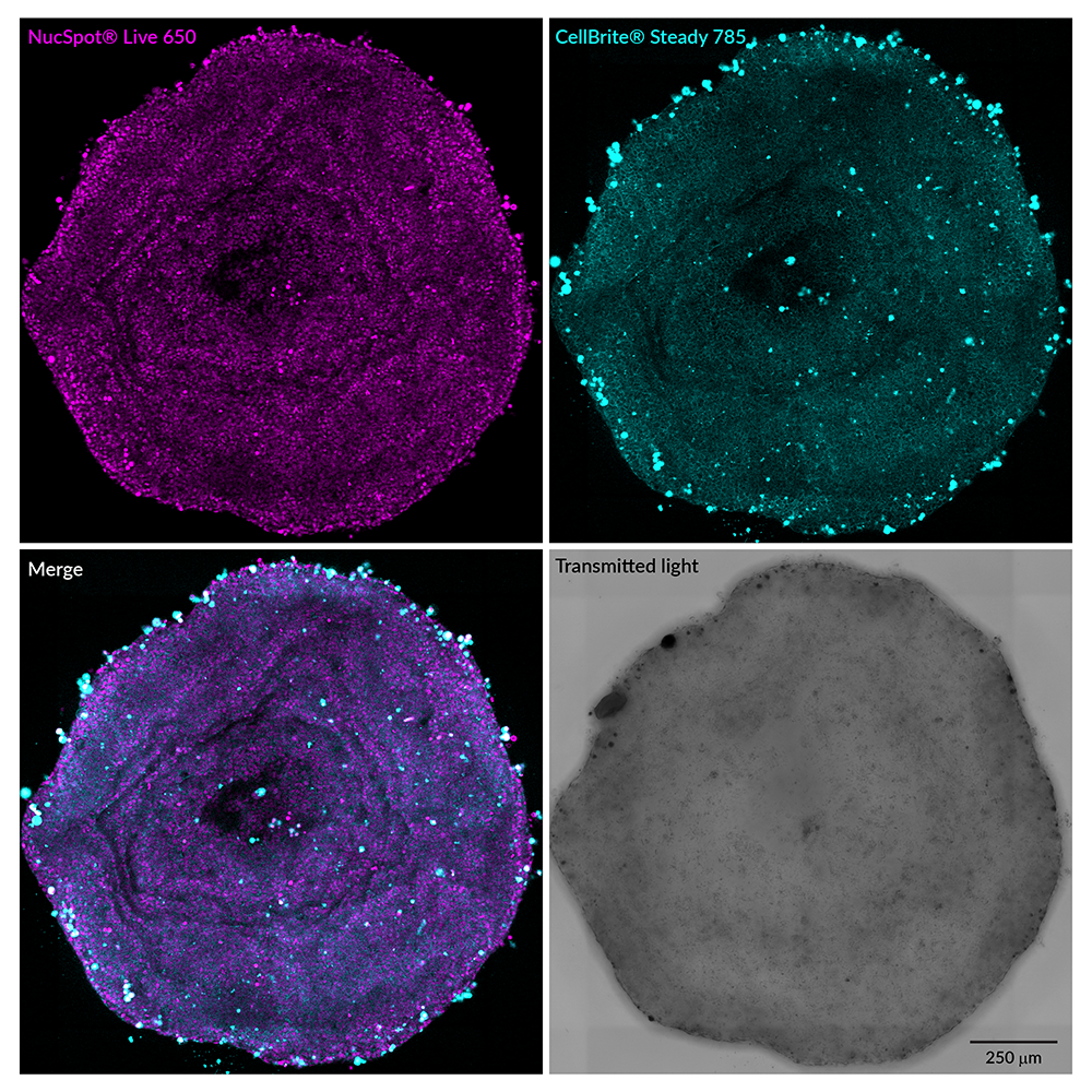 T47-D spheroid stained with NucSpot® Live 650 (nuclei, magenta) and CellBrite® Steady 785 (membranes, cyan). T47-D cells were plated at 20,000 cells per well in a LICORbio 3D Essentials Kit ULA 96-well plate and cultured for 48 hours. The spheroid was stained with dyes in complete medium for 24 hours without a wash step, then imaged on an Evident FV4000 confocal microscope with 20X objective to create a tile-stitched 3D projection image; scale bar: 250 um.
Product Description
NucSpot® Live Cell Nuclear Stains are cell membrane-permeant DNA dyes that specifically stain nuclei in live or fixed cells. They have excellent specificity for DNA without the need for a wash step, with low toxicity for live cell imaging.
Features
- No-wash, nuclear-specific DNA dyes
- Low-toxicity for real-time live cell imaging
- Fix before or after labeling
- NucSpot® Live 650 compatible with SIM, STED, or STORM
- Choose green or far-red fluorescence
Kit Components
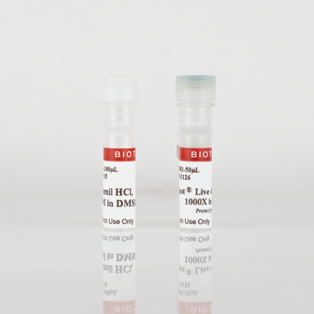
- NucSpot® Live Dye, 1000X in DMSO
- Verapamil, 100 mM in DMSO
Spectral Properties
- NucSpot® Live 488 Ex/Em: 503/518 nm
- NucSpot® Live 650 Ex/Em: 655/681 nm
NucSpot® Live 488 stains nuclei with green fluorescence for the FITC channel, while NucSpot® Live 650 has far-red fluorescence for the Cy®5 channel. NucSpot® Live 650 dye also is compatible with super-resolution imaging by SIM or STED, and has been validated in STORM (Ref. 4). The dyes are supplied as 1000X stock solutions in DMSO, and are supplied with a vial of the efflux pump inhibitor verapamil for optional use, which may increase probe retention and live cell staining in some cell types. Note: NucSpot® Live 488 and NucSpot® Live 650 have dim blue fluorescence in the DAPI channel, and may not be suitable for multicolor imaging with blue probes. NucSpot® Live 650 has also been validated for live imaging of 3D organoid cultures (see figure below). NucSpot® Live Cell Nuclear Stains can be used to stain live gram-positive bacteria, but do not stain live gram-negative bacteria or yeast. See our Cellular Stains Table for more information on how our dyes stain various organisms.
Robust Long-Term Imaging of Nuclei in Live Cells
Figure 1. Time course of live MCF-7 cell staining with NucSpot® Live 488, with or without verapamil. Dye and verapamil were added at time 0 and incubated with cells continuously for three days. Verapamil improves dye retention and staining. NucSpot® Live 488 with verapamil showed no obvious toxicity in MCF-7 cells over the course of 72 hours. With longer incubation times, some extranuclear staining can be observed.
Validated for Live Imaging of Organoids & Other 3D Cultures
Figure 2. 3D tissue dynamics of branching mouse mammary gland organoids. Organoids were incubated with NucSpot® Live 650 and treated with growth factors to trigger post-natal remodeling. The organoids were imaged for 48 hours on the Leica Viventis SCAPE light sheet microscope. Credit: Ayushi Bhanushali, and Sergio P. Acebrón.
Biotium also offers NucSpot® Nuclear Stains for fixed cells in a wide selection of colors from green to near-IR. Also see our wide selection of other Membrane and Organelle Stains, or download the Cellular Stains Brochure.
Cy Dye is a registered trademark of Cytiva.
Technology


 Powered by Bioz