

NucView® 488 and MitoView™ 633 Apoptosis Assay Kit (100 assays)

Cat# 30062
Size : 1kit
Marca : Biotium
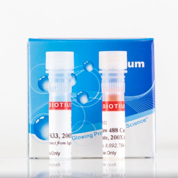
NucView® 488 & MitoView™ 633 Apoptosis Assay Kit
No wash, homogeneous assay for profiling apoptotic cells based on caspase-3 activity and changes in the mitochondrial membrane potential using fluorescence microscopy, real-time imaging, or flow cytometry.
 Source description: Exposure: 000 : 00 : 00 . 274 : 264
Binning: 2 x 2
Gain: 1.000000
%Accumulated%=0
Source description: Exposure: 000 : 00 : 00 . 274 : 264
Binning: 2 x 2
Gain: 1.000000
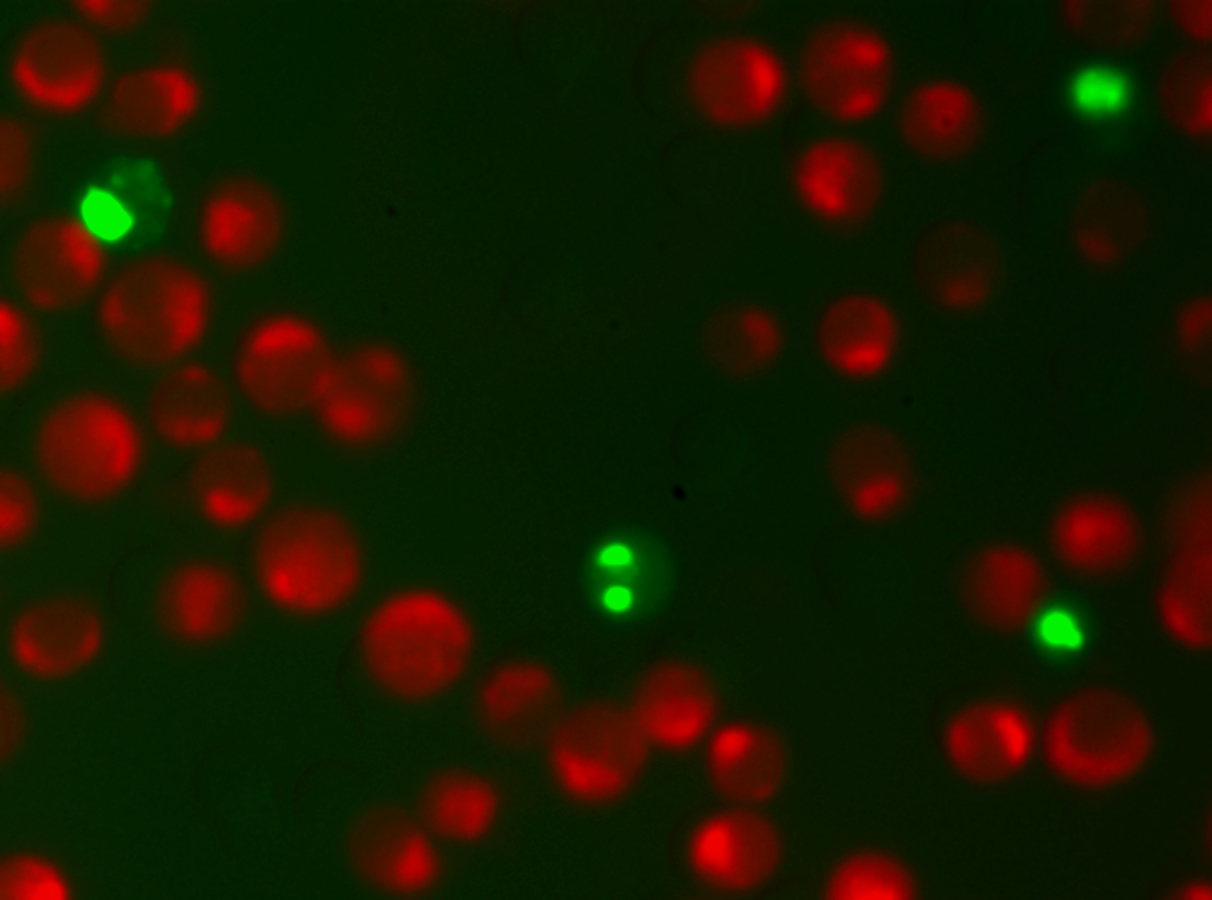
%Accumulated%=0 Click to view GIF animation. HeLa cells were stained with NucView® 488 caspase-3 substrate (2 uM), MitoView™ 633 mitochondrial potential dye (50 nM), and Hoechst 33342 blue nuclear stain (1 ug/mL), then treated with 1 uM staurosporine to induce apoptosis.
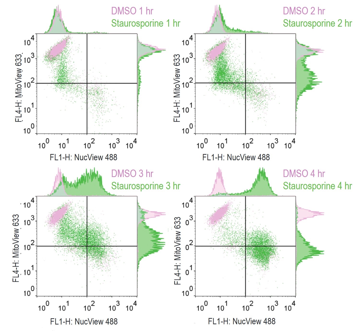 Flow cytometry analysis of Jurkat cells treated with staurosporine (green) to induce
apoptosis, or DMSO controls (pink), using the NucView® 488 and MitoView™ 633 Apoptosis Kit.
Fluorescence was analyzed on a BD FACSCalibur flow cytometer. As apoptosis progresses over
time in staurosporine-treated cells, NucView® 488 signal (FL1, x-axis) increases and mitochondrial
membrane potential measured by MitoView™ 633 staining (FL4, y-axis) decreases.
Flow cytometry analysis of Jurkat cells treated with staurosporine (green) to induce
apoptosis, or DMSO controls (pink), using the NucView® 488 and MitoView™ 633 Apoptosis Kit.
Fluorescence was analyzed on a BD FACSCalibur flow cytometer. As apoptosis progresses over
time in staurosporine-treated cells, NucView® 488 signal (FL1, x-axis) increases and mitochondrial
membrane potential measured by MitoView™ 633 staining (FL4, y-axis) decreases.Product Description
This kit contains NucView® 488 Caspase-3 Substrate to detect apoptotic cells and MitoView™ 633 mitochondrial membrane potential dye.
- Rapid, no-wash, endpoint or real-time assay
- Non-toxic, allowing multi-day experiments to be performed
- For flow cytometry, microscopy or live cell imaging systems
- Dual detection of caspase activity (green) and mitochondrial membrane potential (red/far-red)
NucView® 488 Caspase-3 Substrate is able to detect both intracellular caspase-3 and at the same time stain the cell nucleus, which is known to undergo morphological changes during apoptosis. MitoView™ 633 is a far-red fluorescent mitochondrial dye. The staining is dependent upon the mitochondrial membrane potential; thus, apoptotic cells exhibit a much lower MitoView™ 633 dye staining compared to healthy cells. Furthermore, the spectral separation of these two dyes minimizes fluorescence overlap. See a video of NucView® & MitoView™ in real-time imaging.
Note: The optimal detection settings for MitoView™ 633 are the same as for Cy®5 and other far-red dyes. However, the dye also has visible red fluorescence and can be imaged using Cy®3 settings as well. As a consequence, the dye cannot be used for two-color imaging with other red probes.
To learn about the advantages of monitoring apoptosis using NucView® caspase-3 substrates, visit the NucView® Technology Page.
Technology

The Full Spectrum Blog

Product Attributes
Size
Apoptosis/viability marker
For live or fixed cells
Detection method/readout
Assay type/options
Substrate specificity
Colors
Storage Conditions
Documents, Protocols, SDS and COA

COA
Request the Certificate of AnalysisReferences
Download list of curated NucView® references and validated cell lines.
Citations
Download list of curated NucView® references and validated cell lines.
 Powered by Bioz