

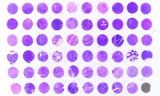
Frozen tissue arrays, also referred to as frozen tissue microarrays (frozen TMA) or cryoarrays, consist of multiple frozen tissue specimens or tissue cores arranged within a common frozen recipient block, commonly using optimal cutting temperature (OCT) compound, followed by cryostat sectioning. This format allows multiple specimens to be analyzed on the same slide under closely matched experimental conditions and can help reduce inter-slide and processing-related variability. Peer-reviewed studies have demonstrated applications of frozen tissue arrays for DNA, RNA, and protein analysis, including immunohistochemistry (IHC), immunocytochemistry, RNA in situ hybridization, and fluorescence in situ hybridization (FISH).
Frozen Tissue Arrays Can Be Organized As:
-
Same-Species Tissue Arrays: Specimens from a single species, allowing comparisons among tissues, anatomical regions, pathological states, experimental groups, or individual samples.
-
Multi-Species Tissue Arrays: Specimens from different species assembled within the same array, supporting comparative tissue analysis and antibody or biomarker studies when appropriate assay validation is performed.
Key Features
-
Frozen Tissue Specimens : Suitable for cryostat sectioning.
-
Multiple Specimens Per Array : Represents multiple specimens on a single array.
-
Preservation of Selected Molecular Targets : Retains selected endogenous molecular targets prior to assay-specific fixation.
-
Standardized Slide-Based Analysis : Enables closely matched analysis conditions across specimens.
-
Broad Research Compatibility : Compatible with histological and molecular research workflows.
Usual Research Applications
-
Protein-expression analysis and immunohistochemistry.
-
Immunofluorescence, RNA in situ hybridization, and DNA analysis.
-
Fluorescence in situ hybridization (FISH).
-
Biomarker screening and comparative tissue analysis.
-
Translational research.