

Rabbit Anti-DHCR7 (C-term) Polyclonal Antibody

Referentie AP11452b-ev
Formaat : 80ul
Merk : Abcepta
DHCR7 Antibody (C-term)
Affinity Purified Rabbit Polyclonal Antibody (Pab)
-
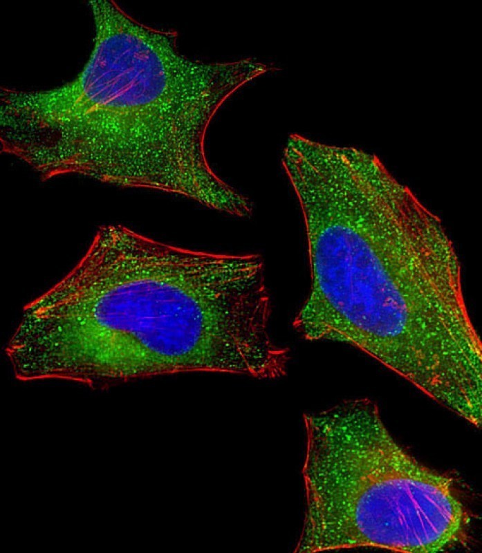
Immunofluorescent analysis of 4% paraformaldehyde-fixed, 0. 1% Triton X-100 permeabilized HeLa (human cervical epithelial adenocarcinoma cell line) cells labeling Pdx1 with AP11452B at 1/25 dilution, followed by Dylight® 488-conjugated goat anti-rabbit IgG (NK179883) secondary antibody at 1/200 dilution (green). Immunofluorescence image showing cytoplasm staining on HeLa cell line. Cytoplasmic actin is detected with Dylight® 554 Phalloidin (PD18466410) at 1/100 dilution (red). The nuclear counter stain is DAPI (blue).

-
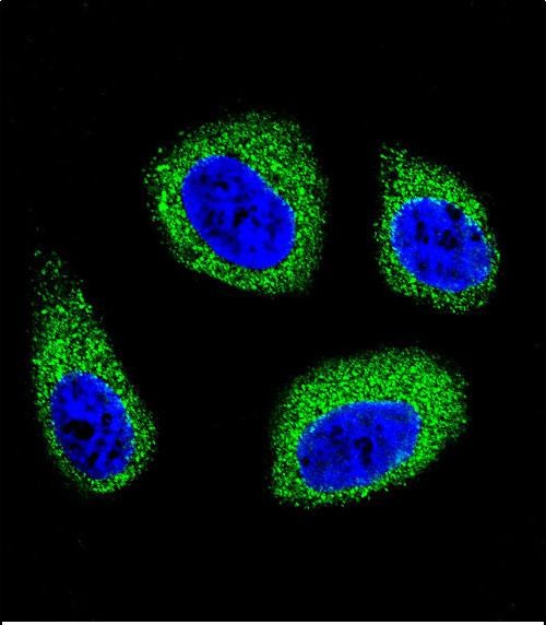
Confocal immunofluorescent analysis of DHCR7 Antibody (C-term)(Cat#AP11452b) with MCF-7 cell followed by Alexa Fluor 488-conjugated goat anti-rabbit lgG (green).DAPI was used to stain the cell nuclear (blue).
-
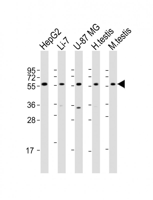
All lanes : Anti-DHCR7 Antibody (C-term) at 1:2000 dilution Lane 1: HepG2 whole cell lysate Lane 2: Li-7 whole cell lysate Lane 3: U-87MG whole cell lysate Lane 4: mouse testis lysate Lysates/proteins at 20 µg per lane. Secondary Goat Anti-Rabbit IgG, (H+L), Peroxidase conjugated at 1/10000 dilution. Predicted band size : 54 kDa Blocking/Dilution buffer: 5% NFDM/TBST.
-
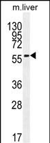
DHCR7 Antibody (C-term) (Cat. #AP11452b) western blot analysis in mouse liver tissue lysates (35ug/lane).This demonstrates the DHCR7 antibody detected the DHCR7 protein (arrow).
-
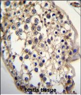
DHCR7 Antibody (C-term) (Cat. #AP11452b)immunohistochemistry analysis in formalin fixed and paraffin embedded human testis tissue followed by peroxidase conjugation of the secondary antibody and DAB staining.This data demonstrates the use of DHCR7 Antibody (C-term) for immunohistochemistry. Clinical relevance has not been evaluated.
-
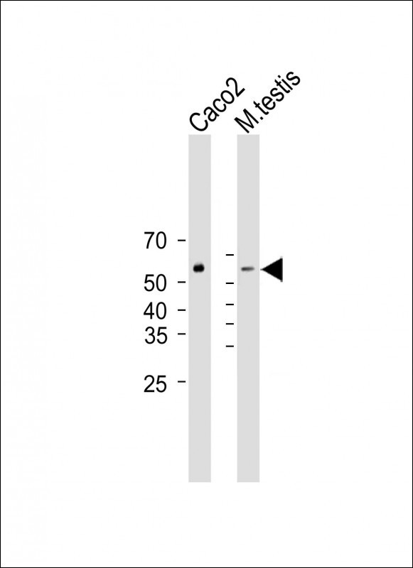
All lanes : Anti-DHCR7 Antibody (C-term) at 1:2000 dilution Lane 1: Caco2 whole cell lysate Lane 2: mouse testis lysate Lysates/proteins at 20 µg per lane. Secondary Goat Anti-Rabbit IgG, (H+L), Peroxidase conjugated (ASP1615) at 1/15000 dilution. Observed band size : 55kDa Blocking/Dilution buffer: 5% NFDM/TBST.
Application 
| WB, IHC-P, IF, E |
|---|---|
| Primary Accession | Q9UBM7 |
| Other Accession | NP_001157289.1, NP_001351.2 |
| Reactivity | Human, Mouse |
| Predicted | Rat, Bovine, Chicken |
| Host | Rabbit |
| Clonality | Polyclonal |
| Isotype | Rabbit IgG |
| Calculated MW | 54489 Da |
| Antigen Region | 437-463 aa |
| Gene ID | 1717 |
|---|---|
| Other Names | 7-dehydrocholesterol reductase, 7-DHC reductase, Putative sterol reductase SR-2, Sterol Delta(7)-reductase, DHCR7, D7SR |
| Target/Specificity | This DHCR7 antibody is generated from rabbits immunized with a KLH conjugated synthetic peptide between 437-463 amino acids from the C-terminal region of human DHCR7. |
| Dilution | WB~~1:2000 IHC-P~~1:50~100 IF~~1:10~50 E~~Use at an assay dependent concentration. |
| Format | Purified polyclonal antibody supplied in PBS with 0.09% (W/V) sodium azide. This antibody is purified through a protein A column, followed by peptide affinity purification. |
| Storage | Maintain refrigerated at 2-8°C for up to 2 weeks. For long term storage store at -20°C in small aliquots to prevent freeze-thaw cycles. |
| Precautions | DHCR7 Antibody (C-term) is for research use only and not for use in diagnostic or therapeutic procedures. |
For Research Use Only. Not For Use In Diagnostic Procedures.
| Name | DHCR7 (HGNC:2860) |
|---|---|
| Synonyms | D7SR |
| Function | Oxidoreductase that catalyzes the last step of the cholesterol synthesis pathway, which transforms cholesta-5,7-dien- 3beta-ol (7-dehydrocholesterol,7-DHC) into cholesterol by reducing the C7-C8 double bond of its sterol core (PubMed:25637936, PubMed:38297129, PubMed:38297130, PubMed:9465114, PubMed:9634533). Can also metabolize cholesta-5,7,24-trien-3beta-ol (7-dehydrodemosterol, 7-DHD) to desmosterol, which is then metabolized by the Delta(24)-sterol reductase (DHCR24) to cholesterol (By similarity). Modulates ferroptosis (a form of regulated cell death driven by iron-dependent lipid peroxidation) through the metabolic breakdown of the anti- ferroptotic metabolites 7-DHC and 7-DHD which, when accumulated, divert the propagation of peroxyl radical-mediated damage from phospholipid components to its sterol core, protecting plasma and mitochondrial membranes from phospholipid autoxidation (PubMed:38297129, PubMed:38297130). |
| Cellular Location | Endoplasmic reticulum membrane; Multi-pass membrane protein |
| Tissue Location | Widely expressed. Most abundant in adrenal gland, liver, testis, and brain. |
- Primary Antibodies
- Cancer
- Cardiovascular
- Metabolism
- Signal Transduction

Provided below are standard protocols that you may find useful for product applications.
Background
This gene encodes an enzyme that removes the C(7-8) double bond in the B ring of sterols and catalyzes the conversion of 7-dehydrocholesterol to cholesterol. This gene is ubiquitously expressed and its transmembrane protein localizes to the endoplasmic reticulum membrane and nuclear outer membrane. Mutations in this gene cause Smith-Lemli-Opitz syndrome (SLOS); a syndrome that is metabolically characterized by reduced serum cholesterol levels and elevated serum 7-dehydrocholesterol levels and phenotypically characterized by mental retardation, facial dysmorphism, syndactyly of second and third toes, and holoprosencephaly in severe cases to minimal physical abnormalities and near-normal intelligence in mild cases. Alternative splicing results in multiple transcript variants that encode the same protein.