

Concanavalin A, CF®680 Conjugate

Referentie 29020
Formaat : 5x1MG
Merk : Biotium
Concanavalin A (Con A) CF® Dye Conjugates
A lectin labeled with our superior fluorescent CF® Dyes is an effective and widely used cell surface stain for yeast, fungi, and mammalian cells.
 Live HeLa cells stained with 50 ug/mL CF®488A ConA (green) and 1 ug/mL Hoechst 33342 (blue) for 10 minutes at 37°C in HBSS, then rinsed and imaged in HBSS.
 Live HeLa cells stained with 50 ug/mL CF®640R ConA (magenta) and 1 ug/mL Hoechst 33342 (blue) for 10 minutes at 37°C in HBSS, then rinsed and imaged in HBSS.
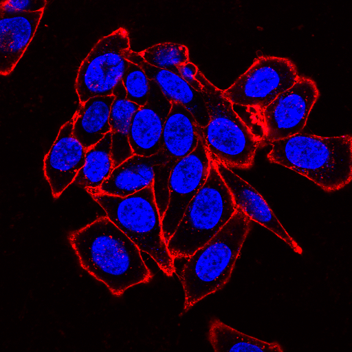 Live HeLa cells stained with 50 ug/mL CF®594 Concanavalin A (red) in HBSS for 10 minutes at 37°C. Nuclei are counterstained with Hoechst (blue).
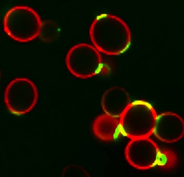 S. cerevisiae stained with CF®488A WGA (green) to stain bud scars and CF®594 ConA (red) to stain the cell wall.
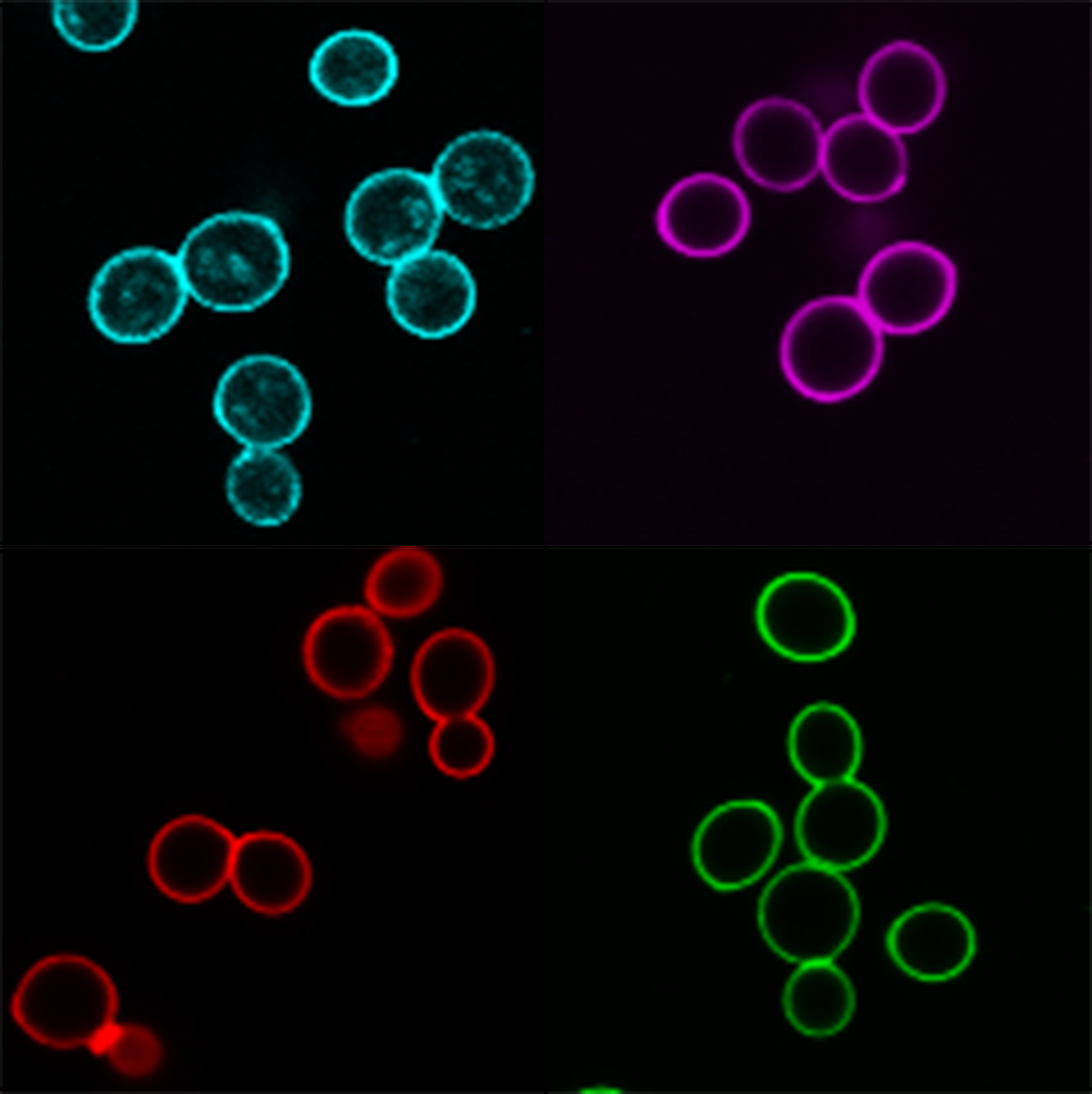
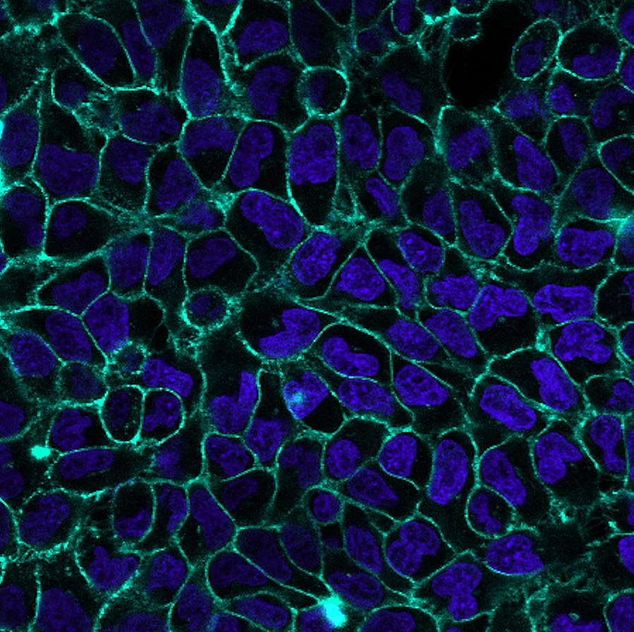 Live HeLa cells were stained for 10 minutes at 37°C with 50 ug/mL CF®748 ConA (cyan) and 1 ug/mL Hoechst (nuclei, blue) in complete culture medium, then rinsed in medium. Fluorescence was imaged with a 40X objective on an Evident FV4000 confocal microscope with 405 nm and 730 nm excitation.
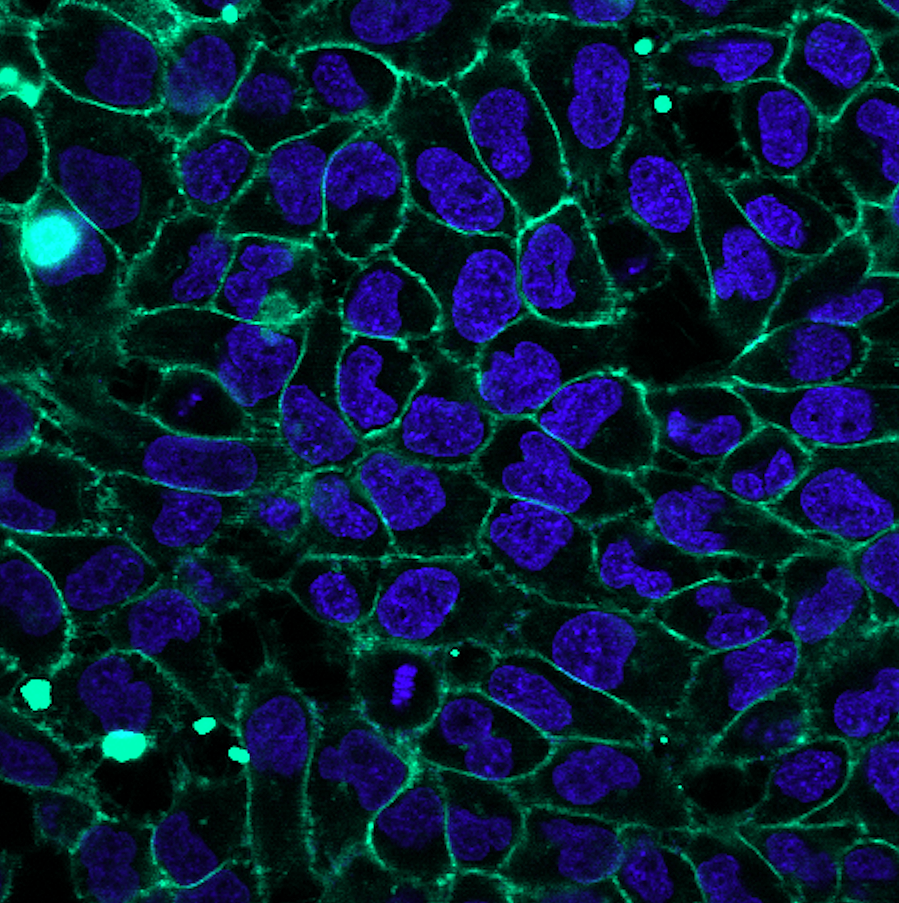 Live HeLa cells were stained for 10 minutes at 37°C with 50 ug/mL CF®790 ConA (cyan) and 1 ug/mL Hoechst (nuclei, blue) in complete culture medium, then rinsed in medium. Fluorescence was imaged with a 40X objective on an Evident FV4000 confocal microscope with 405 nm and 785 nm excitation.
Product Description
Concanavalin A (Con A) is a widely used lectin that selectively binds to the glycoproteins, a-mannopyranosyl and a-glucopyranosyl. These are commonly found in the cell wall of yeast and fungi, and the cell membrane of mammalian cells and tissues.
- Stain the cell wall of yeast and fungi, and the cell membrane of mammalian cells and tissues
- Detect glycoconjugates in microscopy and flow cytometry
- Stain glycoproteins in gels
- Withstands fixation and permeabilization
- Choice of 13 CF® Dyes from UV to near-infrared
- Superior CF® Dyes are bright, photostable, and water-soluble
Lectins are also versatile probes for detecting glycoconjugates in microscopy and flow cytometric applications and for gel staining of glycoproteins. In neutral and alkaline solutions, Con A exists as a tetramer with a molecular weight of approximately 104 kDa. In acidic solutions (pH below 5.0), Con A exists as a dimer. Con A can be used to selectively stain the cell surface of live cells, and withstand fixation and permeabilization. When cells are fixed and permeabilized before staining, fluorescent lectins stain both cell surface and organelles in the secretory pathway.
Find the Right Stain for Your Application
Con A and other lectins are carbohydrate binding proteins that recognize specific sugar moieties on glycoproteins. The presence and distribution of these targets vary between cell types and tissues. As a result, other cell surface stains or other lectin conjugates, Wheat Germ Agglutinin (WGA) Conjugates and PNA Lectin Conjugates, may produce better surface staining and may be more appropriate for your cell type. Lectin conjugates can be used to selectively stain the cell surface of live cells, and withstand fixation and permeabilization. When cells are fixed and permeabilized before staining, fluorescent lectins stain both cell surface and organelles in the secretory pathway. Lectins may be toxic or stimulatory to live cells depending on cell type. To find the right stain for your application, see our Membrane & Cell Surface Stains Comparison, or download our Membrane & Surface Stains Brochure. See our Cellular Stains Table for more information on how our dyes stain various organisms.
Superior CF® Dyes
Biotium’s next-generation CF® Dyes were designed to be highly water-soluble with advantages in brightness and photostability compared to Alexa Fluor®, DyLight®, and other fluorescent dyes. Learn more about CF® Dyes.
Note: Conjugates of blue-fluorescent dyes like CF®350, CF®405S and CF®405M are not recommended for detecting low abundance targets and may be challenging to use in tissue specimens. Blue dyes have lower fluorescence and photostability, and cells and tissue have high autofluorescence in blue wavelengths, resulting in lower signal to noise compared to other colors.
Concanavalin A Conjugates
| Conjugation | Ex/Em | Size | Catalog No. | Dye Features |
|---|---|---|---|---|
| CF®350 | 355/450 nm | 5 X 1 mg | 29015 | CF®350 Features |
| CF®405S | 411/431 nm | 5 X 1 mg | 29075 | CF®405S Features |
| CF®405M | 416/452 nm | 5 X 1 mg | 29074 | CF®405M Features |
| CF®488A | 490/516 nm | 5 X 1 mg | 29016 | CF®488A Features |
| CF®594 | 593/615 nm | 5 X 1 mg | 29017 | CF®594 Features |
| CF®633 | 629/650 nm | 5 X 1 mg | 29018 | CF®633 Features |
| CF®640R | 642/663 nm | 5 X 1 mg | 29019 | CF®640R Features |
| CF®680 | 681/698 nm | 5 X 1 mg | 29020 | CF®680 Features |
| CF®740 | 742/767 nm | 5 X 1 mg | 29136 | CF®740 Features |
| CF®748 | 747/770 nm | 5 X 1 mg | 29141 | CF®748 Features |
| CF®750 | 755/779 nm | 5 X 1 mg | 29080 | CF®750 Features |
| CF®770 | 770/797 nm | 5 X 1 mg | 29058 | CF®770 Features |
| CF®790 | 783/808 nm | 5 X 1 mg | 29142 | CF®790 Features |
Full List of Lectin Conjugates
| Product | Features |
|---|---|
| CF® Dye Concanavalin A (Con A) | • Cell surface stain for yeast, fungi, and mammalian cells • Selectively binds to a-mannopyranosyl and a-glucopyranosy residues • Available with a wide selection of CF® Dyes |
| CF® Dye Wheat Germ Agglutinin (WGA) | • Cell surface stain for mammalian cells and gram+ bacteria • Also stains yeast bud scars • Has high affinity for sialic acid and N-acetylglucosamine • Choose from a wide selection of CF® Dyes or HRP |
| CF® Dye Peanut Lectin (PNA) from Arachis hypogaea | • Specific for terminal β-galactose and binds preferentially to galactosyl (β-1,3) N-acetylgalactosamine • Choice of 6 CF® dye colors |
| CF® Dye Lycopersicon Esculentum (Tomato) Lectin (LEL, TL) | • Marker for blood vessels and microglial cells • Binds to [GlcNAc] 1,3-N-acetylglucosamine, glycophorin, and Tamm-Horsfall glycoprotein • Used to study tumor angiogenesis or tracing neovascular development in xenograft models • Choice of 7 CF® Dyes or biotin |
| CF® Dye Ulex Europaeus Agglutinin I (UEA I) | • Marker for human endothelial cells and incompletely differentiated gastrin cells • Binds to glycoproteins and glycolipids containing α-linked fucose residues • Choice of 7 CF® Dyes or biotin |
| CF® Dye Phaseolus Vulgaris Leucoagglutinin (PHA-L) | • Used to stimulate lymphocyte and T cell proliferation • Choice of 7 CF® Dyes or biotin |
| CF® Dye Datura Stramonium Lectin (DSL) | • Binds to (beta-1,4) linked N-acetylglucosamine oligomers • Choice of 7 CF® Dyes or biotin |
| CF® Dye Sambucus Nigra Lectin (SNA, EBL)) | • Binds to sialic acid attached to terminal galactose • Choice of 7 CF® Dyes or biotin |
Technology

Tech Tips

Product Attributes
Size
Probe cellular localization
For live or fixed cells
Cell permeability
Fixation options
Colors
Documents, Protocols, SDS and COA

Protocols
Datasheet
COA
Request the Certificate of AnalysisReferences
CF®680 ConA
Acta Histochem (2019), DOI: 10.1016/j.acthis.2018.11.006.
Download a list of CF® dye references.
Citations
CF®680 ConA
Acta Histochem (2019), DOI: 10.1016/j.acthis.2018.11.006.
Download a list of CF® dye references.
 Powered by Bioz