


Dengue virus NS1 protein antibody
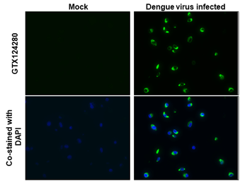
Dengue virus NS1 protein antibody detects Dengue virus NS1 protein protein at cytoplasm by immunohistochemical analysis.Sample: Paraffin-embedded dengue virus BHK-21.Green: Dengue virus NS1 protein stained by Dengue virus NS1 protein antibody (GTX124280) diluted at 1:1000.Blue: Fluoroshield with DAPI (GTX30920).Antigen Retrieval: Citrate buffer, pH 6.0, 15 min
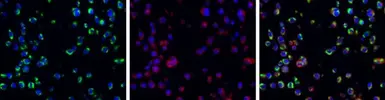
Dengue virus NS1 protein antibody detects Dengue virus NS1 protein protein at cytoplasm by immunohistochemical analysis.Sample: Paraffin-embedded BHK-21 infected with dengue virus.Green: Dengue virus NS1 protein stained by Dengue virus NS1 protein antibody (GTX124280) diluted at 1:2000.Red: alpha Tubulin, a cytoskeleton marker, stained by alpha Tubulin antibody [GT114] (GTX628802) diluted at 1:1000.Blue: Fluoroshield with DAPI (GTX30920).Antigen Retrieval: Citrate buffer, pH 6.0, 15 min
The results may vary from different DENV strains, viral infection status, and culture conditions.
Reference: Proc Natl Acad Sci U S A. 2013 Apr 23;110(17):6795-9, J Infect Dis. 2011 Jun 15;203(12):1775-83.
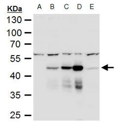
NS1 (Dengue virus) antibody detects NS1 (Dengue virus) protein by western blot analysis.
A. 66 μg BHK-21 whole cell extract
B. 66 μg whole cell extract of Dengue virus type 1 infected BHK-21 cells
C. 20 μg whole cell extract of Dengue virus type 2 infected BHK-21 cells
D. 10 μg whole cell extract of Dengue virus type 3 infected BHK-21 cells
E. 66 μg whole cell extract of Dengue virus type 4 infected BHK-21 cells
10% SDS-PAGE
NS1 (Dengue virus) antibody (GTX124280) dilution: 1:3000
The HRP-conjugated anti-rabbit IgG antibody (GTX213110-01) was used to detect the primary antibody.
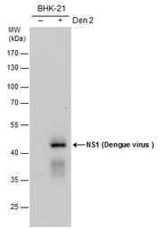
NS1 (Dengue virus ) antibody detects NS1 (Dengue virus ) protein by Western blot analysis. Un-infected (-) and infected (+, DENV2 infection for ) BHK-21 whole cell extracts (20 μg) were separated by 10% SDS-PAGE, and the membrane was blotted with NS1 (Dengue virus ) antibody (GTX124280) diluted by 1:2000.
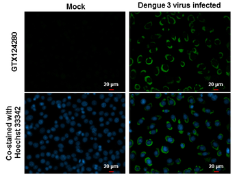
NS1 (Dengue virus) antibody detects NS1 (Dengue virus) protein at cytoplasm by immunofluorescent analysis.
Samples: BHK-21 cells mock (left) and infected with Dengue virus 3 (right) were fixed in MeOH.
Green: NS1 (Dengue virus) protein stained by NS1 (Dengue virus) antibody (GTX124280) diluted at 1:2000.
Blue: Hoechst 33342 staining.
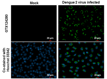
NS1 (Dengue virus) antibody detects NS1 (Dengue virus) protein at cytoplasm by immunofluorescent analysis.
Samples: BHK-21 cells mock (left) and infected with Dengue virus 2 (right) were fixed in MeOH.
Green: NS1 (Dengue virus) protein stained by NS1 (Dengue virus) antibody (GTX124280) diluted at 1:2000.
Blue: Hoechst 33342 staining.
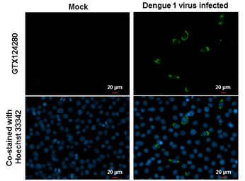
NS1 (Dengue virus) antibody detects NS1 (Dengue virus) protein at cytoplasm by immunofluorescent analysis.
Samples: BHK-21 cells mock (left) and infected with Dengue virus 1 (right) were fixed in MeOH.
Green: NS1 (Dengue virus) protein stained by NS1 (Dengue virus) antibody (GTX124280) diluted at 1:2000.
Blue: Hoechst 33342 staining.
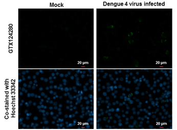
NS1 (Dengue virus) antibody detects NS1 (Dengue virus) protein at cytoplasm by immunofluorescent analysis.
Samples: BHK-21 cells mock (left) and infected with Dengue virus 4 (right) were fixed in MeOH.
Green: NS1 (Dengue virus) protein stained by NS1 (Dengue virus) antibody (GTX124280) diluted at 1:2000.
Blue: Hoechst 33342 staining.
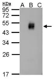
Sample (20 μg of whole cell lysate)
A: BHK-21
B: Dengue virus 2 infect BHK-21
C: JEV infect BHK-21
12% SDS PAGE
GTX124280 diluted at 1:2000
The HRP-conjugated anti-rabbit IgG antibody (GTX213110-01) was used to detect the primary antibody.