

Capsid protein VP2 Recombinant Protein

Référence OPCA02150-20UG
Conditionnement : 20ug
Marque : Aviva Systems Biology
Capsid protein VP2 Recombinant Protein (isolate AU) (OPCA02150)
| Datasheets/Manuals | Printable datasheet for Capsid protein VP2 Recombinant Protein (isolate AU) (OPCA02150) |
|---|
| Predicted Species Reactivity | Human parvovirus B19|Human Parvovirus B19 |
|---|---|
| Product Format | Liquid or Lyophilized powder |
| Host | Human parvovirus B19 |
| Additional Information | Relevance: Capsid protein self-assbles to form an icosahedral capsid with a T=1 symmetry, about 22 nm in diameter, and consisting of 60 copies of two size variants of the capsid proteins, VP1 and VP2, which differ by the presence of an N-terminal extension in the minor protein VP1. The capsid encapsulates the genomic ssDNA. Capsid proteins are responsible for the attachment to host cell receptors, such as the glycosphingolipid globoside or the integrin heterodimer ITGAV/ITGB1. This attachment induces virion internalization predominantly through clathrin-dependent endocytosis. Binding to the host receptors also induces capsid rearrangents leading to surface exposure of VP1 N-terminus, specifically its phospholipase A2-like region and nuclear localization signal(s). VP1 N-terminus might serve as a lipolytic enzyme to breach the endosomal mbrane during entry into host cell. Intracytoplasmic transport involves microtubules and interaction between capsid proteins and host dynein. Exposure of nuclear localization signal probably allows nuclear import of capsids . |
| Reconstitution and Storage | -20°C or -80°C |
| Formulation | 20 mM Tris-HCl based buffer, pH 8.0 |
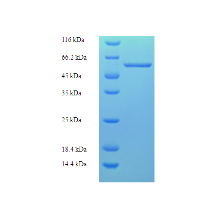
| Purity | Greater than 90% as determined by SDS-PAGE. |
| Peptide Sequence | MTSVNSAEASTGAGGGGSNSVKSMWSEGATFSANSVTCTFSRQFLIPYDPEHHYKVFSPAASSCHNASGKEAKVCTISPIMGYSTPWRYLDFNALNLFFSPLEFQHLIENYGSIAPDALTVTISEIAVKDVTDKTGGGVQVTDSTTGRLCMLVDHEYKYPYVLGQGQDTLAPELPIWVYFPPQYAYLTVGDVNTQGISGDSKKLASEESAFYVLEHSSFQLLGTGGTASMSYKFPPVPPENLEGCSQHFYEMYNPLYGSRLGVPDTLGGDPKFRSLTHEDHAIQPQNFMPGPLVNSVSTKEGDSSNTGAGKALTGLSTGTSQNTRISLRPGPVSQPYHHWDTDKYVTGINAISHGQTTYGNAEDKEYQQGVGRFPNEKEQLKQLQGLNMHTYFPNKGTQQYTDQIERPLMVGSVWNRRALHYESQLWSKIPNLDDSFKTQFAALGGWGLHQPPPQIFLKILPQSGPIGGIKSMGITTLVQYAVGIMTVTMTFKLGPRKATGRWNPQPGVYPPHAAGHLPYVLYDPTATDAKQHHRHGYEKPEELWTAKSRVHPL |
| Protein Sequence | MTSVNSAEASTGAGGGGSNSVKSMWSEGATFSANSVTCTFSRQFLIPYDPEHHYKVFSPAASSCHNASGKEAKVCTISPIMGYSTPWRYLDFNALNLFFSPLEFQHLIENYGSIAPDALTVTISEIAVKDVTDKTGGGVQVTDSTTGRLCMLVDHEYKYPYVLGQGQDTLAPELPIWVYFPPQYAYLTVGDVNTQGISGDSKKLASEESAFYVLEHSSFQLLGTGGTASMSYKFPPVPPENLEGCSQHFYEMYNPLYGSRLGVPDTLGGDPKFRSLTHEDHAIQPQNFMPGPLVNSVSTKEGDSSNTGAGKALTGLSTGTSQNTRISLRPGPVSQPYHHWDTDKYVTGINAISHGQTTYGNAEDKEYQQGVGRFPNEKEQLKQLQGLNMHTYFPNKGTQQYTDQIERPLMVGSVWNRRALHYESQLWSKIPNLDDSFKTQFAALGGWGLHQPPPQIFLKILPQSGPIGGIKSMGITTLVQYAVGIMTVTMTFKLGPRKATGRWNPQPGVYPPHAAGHLPYVLYDPTATDAKQHHRHGYEKPEELWTAKSRVHPL |
| Storage Buffer | If the delivery form is liquid, the default storage buffer is Tris/PBS-based buffer, 5%-50% glycerol. If the delivery form is lyophilized powder, the buffer before lyophilization is Tris/PBS-based buffer, 6% Trehalose, pH 8.0. |
| Source | E.coli |
| Protein Range | 228-781 aa |
| Tag | N-terminal 6xHis-tagged |
| Reference | Nucleotide sequence and genome organization of human parvovirus B19 isolated from the serum of a child during aplastic crisis.Shade R.O., Blundell M.C., Cotmore S.F., Tattersall P., Astell C.R.J. Virol. 58:921-936(1986) |
|---|---|
| Alias Symbols | Coat protein VP1. |
| Protein Name | Capsid protein VP1 |
| Description of Target | Capsid protein self-assembles to form an icosahedral capsid with a T=1 symmetry, about 22 nm in diameter, and consisting of 60 copies of two size variants of the capsid proteins, VP1 and VP2, which differ by the presence of an N-terminal extension in the minor protein VP1. The capsid encapsulates the genomic ssDNA. Capsid proteins are responsible for the attachment to host cell receptors, such as the glycosphingolipid globoside or the integrin heterodimer ITGAV/ITGB1. This attachment induces virion internalization predominantly through clathrin-dependent endocytosis. Binding to the host receptors also induces capsid rearrangements leading to surface exposure of VP1 N-terminus, specifically its phospholipase A2-like region and nuclear localization signal(s). VP1 N-terminus might serve as a lipolytic enzyme to breach the endosomal membrane during entry into host cell. Intracytoplasmic transport involves microtubules and interaction between capsid proteins and host dynein. Exposure of nuclear localization signal probably allows nuclear import of capsids (By similarity). |
| Uniprot ID | P07299 |
| Protein Size (# AA) | Partial |
| Molecular Weight | 64.8 kDa |