


Product Info Summary
| SKU: | PB9771 |
|---|---|
| Size: | 100 μg/vial |
| Reactive Species: | Human, Mouse, Rat |
| Host: | Rabbit |
| Application: | IHC, WB |
Customers Who Bought This Also Bought
Cytoplasmic and Nuclear Protein Extraction Kit (AR0106)
Product info
Product Name
Anti-PDGF Receptor alpha/PDGFRA Antibody Picoband®
SKU/Catalog Number
PB9771
PB0822 is an alternative SKU for this antibody, used in previous lots.
Size
100 μg/vial
Form
Lyophilized
Description
Boster Bio Anti-PDGF Receptor alpha/PDGFRA Antibody Picoband® catalog # PB9771. Tested in IHC, WB applications. This antibody reacts with Human, Mouse, Rat. The brand Picoband indicates this is a premium antibody that guarantees superior quality, high affinity, and strong signals with minimal background in Western blot applications. Only our best-performing antibodies are designated as Picoband, ensuring unmatched performance.
Storage & Handling
Store at -20˚C for one year from date of receipt. After reconstitution, at 4˚C for one month. It can also be aliquotted and stored frozen at -20˚C for six months. Avoid repeated freeze-thaw cycles.
Cite This Product
Anti-PDGF Receptor alpha/PDGFRA Antibody Picoband® (Boster Biological Technology, Pleasanton CA, USA, Catalog # PB9771)
Host
Rabbit
Contents
Each vial contains antibody formulated with stabilizing components, 0.9 mg NaCl, 0.2 mg Na2HPO4, and 0.05 mg NaN3.
*This antibody is supplied in a stabilized formulation.
Compatibility with conjugation reactions depends on the chemistry of the conjugation method used.
For conjugation methods that are not compatible with the stabilizing components present in this formulation, a carrier-free antibody format is required.
Clonality
Polyclonal
Isotype
Rabbit IgG
Immunogen
A synthetic peptide corresponding to a sequence at the C-terminus of human PDGFRA, identical to the related mouse sequence, and different from the related rat sequence by one amino acid.
Cross-reactivity
No cross-reactivity with other proteins.
Reactive Species
PB9771 is reactive to PDGFRA in Human, Mouse, Rat
Observed Molecular Weight
123 kDa
Calculated molecular weight
122.7 kDa
Background of PDGFRA
PDGFRA (Platelet-derived growth factor receptor, alpha), also called PDGFR2, encodes a cell surface tyrosine kinase receptor for members of the platelet-derived growth factor family. The PDGFA gene is mapped on 4q12. The PDGFRA-FIP1L1 gene is a constitutively activated tyrosine kinase that transforms hematopoietic cells and is a therapeutic target of imatinib. And the PDGFRA gene contains 23 exons spanning about 65 kb. Using the human PDGFRA promoter linked to a luciferase reporter, Joosten et al. showed that PAX1 acts as a transcriptional activator of the PDGFRA gene in differentiated human embryonal carcinoma cells. PDGFRA is responsible for mediating cellular contraction of multiple growth factors: TGFB1 and members of the PDGF family. Lei et al. noted that in the rabbit model of the disease, PDGFRA is dramatically more capable of promoting PVR than is the closely related PDGFRB. PDGFRA is a critical receptor required for human CMV infection, and thus a target for novel antiviral therapies.
Antibody Validation
Boster validates all antibodies on WB, IHC, ICC, Immunofluorescence, and ELISA with known positive control and negative samples to ensure specificity and high affinity, including thorough antibody incubations.
Application & Images
Applications
PB9771 is guaranteed for IHC, WB Boster Guarantee
Recommend Dilution
| Application | Dilution | Species |
|---|---|---|
| Immunohistochemistry (Paraffin-embedded Section) | 0.5-1μg/ml | Human |
| Western blot | 0.1-0.5μg/ml | Human, Mouse, Rat |
Tested application
Suggested blocking solution with 5% non-fat milk or BSA; (*)Recommended protein loading: 20-40 µg per lane
Use TE buffer pH 9.0 for antigen retrieval; (*) citrate buffer pH 6.0 is an alternative.
Validation Images & Assay Conditions
Click image to see more details
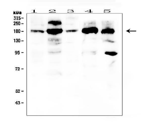
Western blot analysis of PDGFRA using anti-PDGFRA antibody (PB9771).
Electrophoresis was performed on a 5-20% SDS-PAGE gel at 70V (Stacking gel) / 90V (Resolving gel) for 2-3 hours. The sample well of each lane was loaded with 50ug of sample under reducing conditions.
Lane 1: human HT1080 whole cell lysates,
Lane 2: human Caco-2 whole cell lysates,
Lane 3: human U20S whole cell lysates,
Lane 4: human PC-3 whole cell lysates,
Lane 5: human 293T whole cell lysates.
After Electrophoresis, proteins were transferred to a Nitrocellulose membrane at 150mA for 50-90 minutes. Blocked the membrane with 5% Non-fat Milk/ TBS for 1.5 hour at RT. The membrane was incubated with rabbit anti-PDGFRA antigen affinity purified polyclonal antibody (Catalog # PB9771) at 0.5 μg/mL overnight at 4°C, then washed with TBS-0.1%Tween 3 times with 5 minutes each and probed with a goat anti-rabbit IgG-HRP secondary antibody at a dilution of 1:10000 for 1.5 hour at RT. The signal is developed using an Enhanced Chemiluminescent detection (ECL) kit (Catalog # EK1002) with Tanon 5200 system. A specific band was detected for PDGFRA at approximately 123KD. The expected band size for PDGFRA is at 180KD.
Click image to see more details
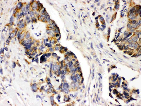
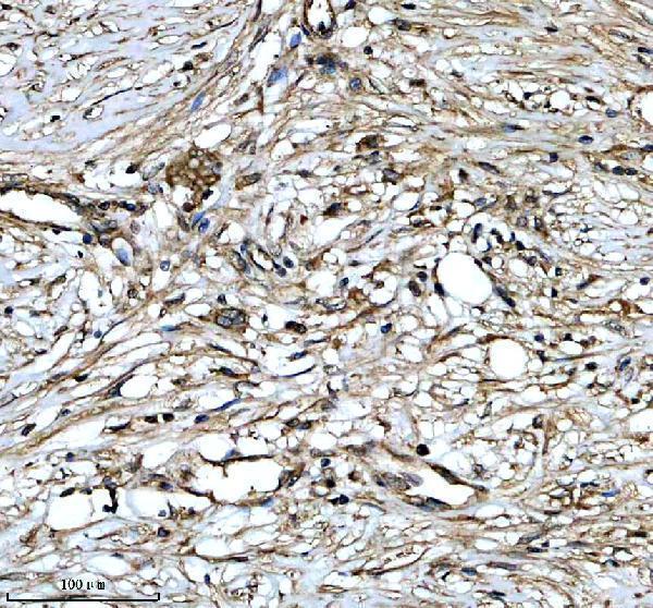
IHC analysis of PDGFRA using anti-PDGFRA antibody (PB9771).
PDGFRA was detected in paraffin-embedded section of Human Intestinal Cancer Tissue. Heat mediated antigen retrieval was performed in citrate buffer (pH6, epitope retrieval solution) for 20 mins. The tissue section was blocked with 10% goat serum. The tissue section was then incubated with 1μg/ml rabbit anti-PDGFRA Antibody (PB9771) overnight at 4°C. Biotinylated goat anti-rabbit IgG was used as secondary antibody and incubated for 30 minutes at 37°C. The tissue section was developed using Strepavidin-Biotin-Complex (SABC)(Catalog # SA1022) with DAB as the chromogen.
Click image to see more details
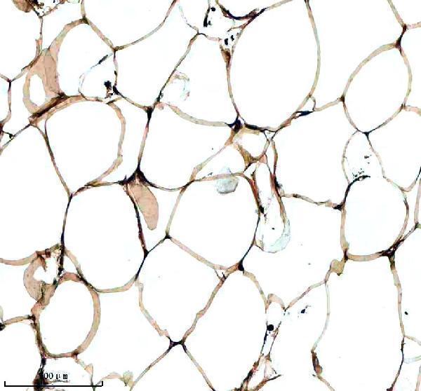
IHC analysis of PDGFRA using anti-PDGFRA antibody (PB9771).
PDGFRA was detected in a paraffin-embedded section of human breast cancer tissue. Heat mediated antigen retrieval was performed in EDTA buffer (pH 8.0, epitope retrieval solution). The tissue section was blocked with 10% goat serum. The tissue section was then incubated with 5 μg/mL rabbit anti-PDGFRA Antibody (PB9771) overnight at 4°C. Peroxidase Conjugated Goat Anti-rabbit IgG was used as secondary antibody and incubated for 30 minutes at 37°C. The tissue section was developed using HRP Conjugated Rabbit IgG Super Vision Assay Kit (Catalog # SV0002) with DAB as the chromogen.
Click image to see more details
IHC analysis of PDGFRA using anti-PDGFRA antibody (PB9771).
PDGFRA was detected in a paraffin-embedded section of human breast cancer tissue. Heat mediated antigen retrieval was performed in EDTA buffer (pH 8.0, epitope retrieval solution). The tissue section was blocked with 10% goat serum. The tissue section was then incubated with 5 μg/mL rabbit anti-PDGFRA Antibody (PB9771) overnight at 4°C. Peroxidase Conjugated Goat Anti-rabbit IgG was used as secondary antibody and incubated for 30 minutes at 37°C. The tissue section was developed using HRP Conjugated Rabbit IgG Super Vision Assay Kit (Catalog # SV0002) with DAB as the chromogen.
Click image to see more details
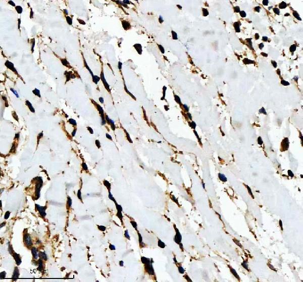
IHC analysis of PDGFRA using anti-PDGFRA antibody (PB9771).
PDGFRA was detected in a paraffin-embedded section of human colon cancer tissue. Heat mediated antigen retrieval was performed in EDTA buffer (pH 8.0, epitope retrieval solution). The tissue section was blocked with 10% goat serum. The tissue section was then incubated with 5 μg/mL rabbit anti-PDGFRA Antibody (PB9771) overnight at 4°C. Peroxidase Conjugated Goat Anti-rabbit IgG was used as secondary antibody and incubated for 30 minutes at 37°C. The tissue section was developed using HRP Conjugated Rabbit IgG Super Vision Assay Kit (Catalog # SV0002) with DAB as the chromogen.
Specific Publications For Anti-PDGF Receptor alpha/PDGFRA Antibody Picoband® (PB9771)
Loading publications
Recommended Resources
Here are featured tools and databases that you might find useful.