

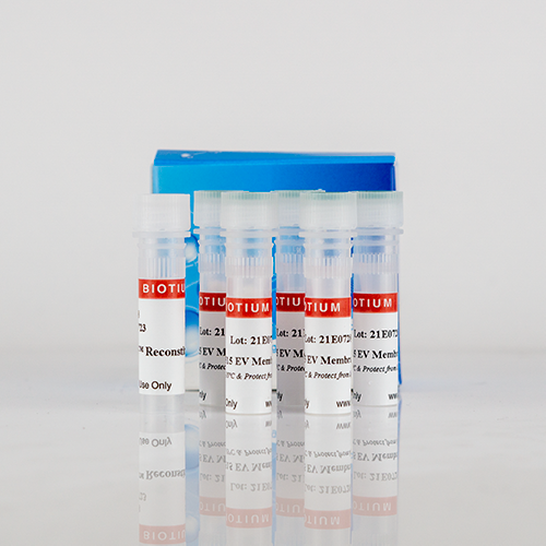
ExoBrite™ 410/450 CTB EV Staining Kit, 500 labelings

Referência 30111
Tamanho : 1kit
Marca : Biotium
ExoBrite™ CTB EV Staining Kits
Fluorescent cholera toxin subunit B (CTB) conjugates that are optimized for bright and clean staining of extracellular vesicles for flow cytometry.
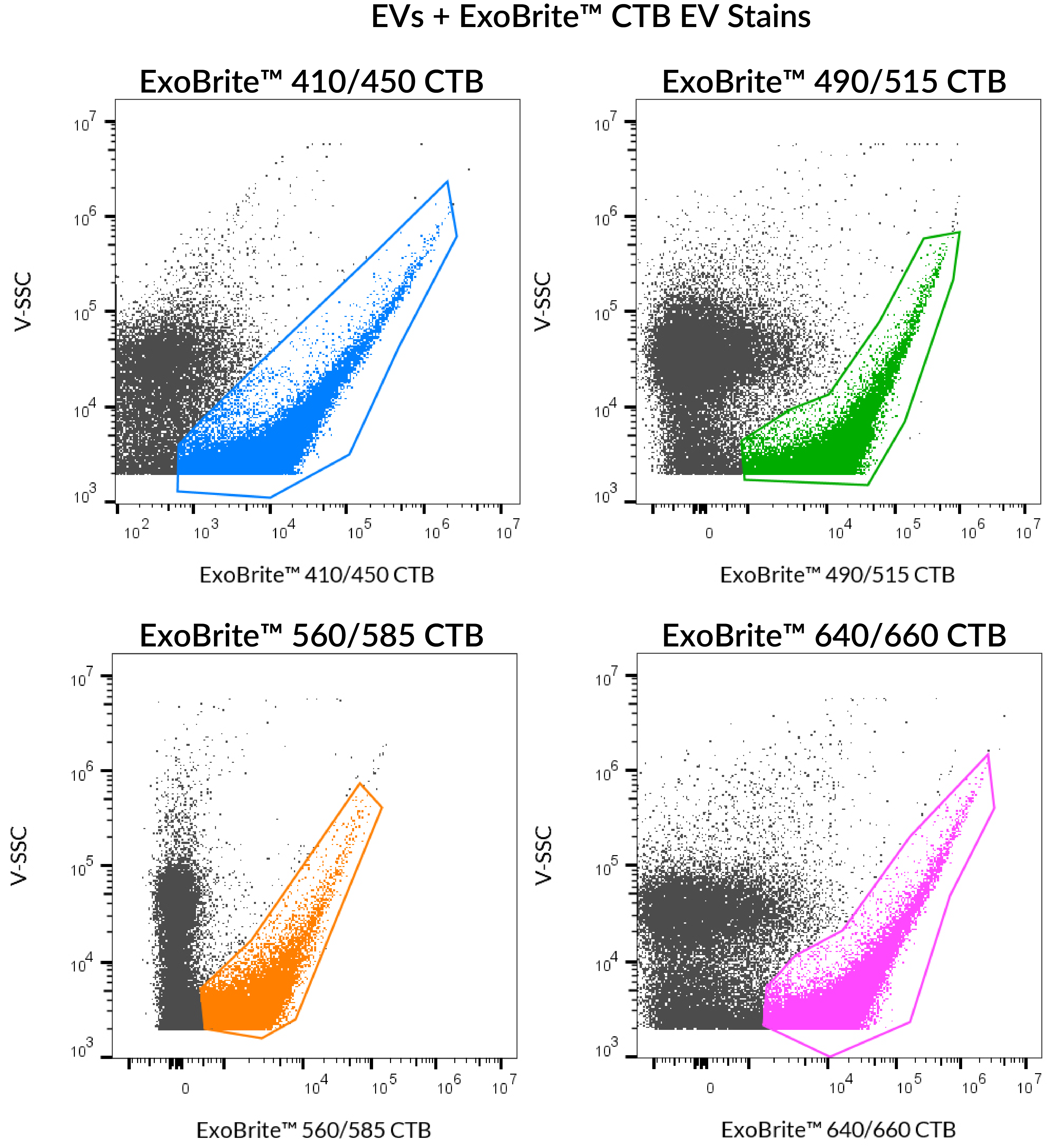 SEC-purified MCF-7-derived exosomes were stained with ExoBrite™ CTB EV Stains. 410/450 was detected in the Pacific Blue channel, 490/515 was detected in the FITC channel, 560/585 was detected in the PE channel, and 640/660 was detected in the APC channel.
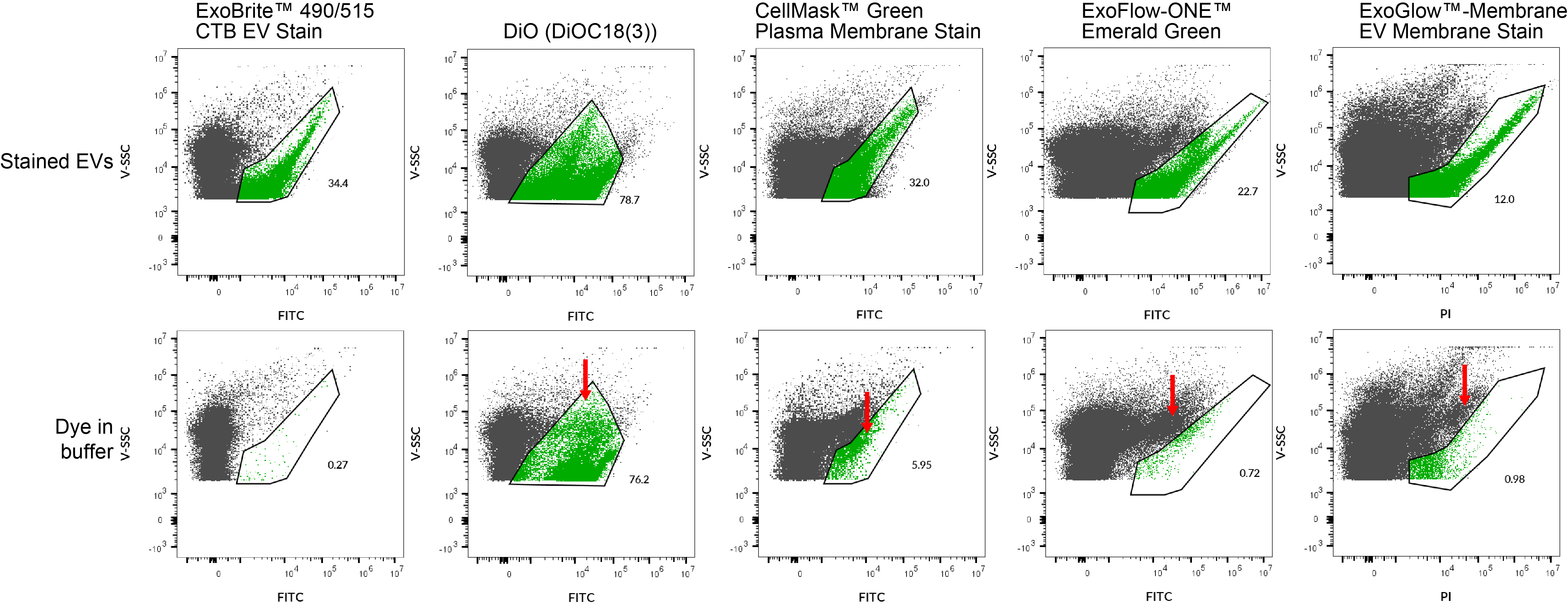 ExoBrite™ CTB EV Stains have less background and more complete staining of extracellular vesicles (EVs) than other classic and competitor dyes. EVs were purified from MCF-7 cell supernatant using size exclusion chromatography (SEC). The purified EVs were stained in PBS with the indicated dyes (top row). The stained EV population was gated, with the number showing the percentage of particles falling within the exosome gate. Each dye was also added to filtered PBS (bottom row), to look for dye aggregation and non-specific background. Red arrows indicate these dye aggregates. Lipophilic dyes like DiO show a high number of particles of similar size to EVs, making them unsuited for small particle staining. CellMask™ also shows an unacceptable amount of dye aggregation falling within the EV gate. The ExoFlow-ONE™ and ExoGlow™ dyes form aggregates that can mostly be gated away from the EVs, but they also show less-complete coverage of EVs than ExoBrite™ CTB stains.
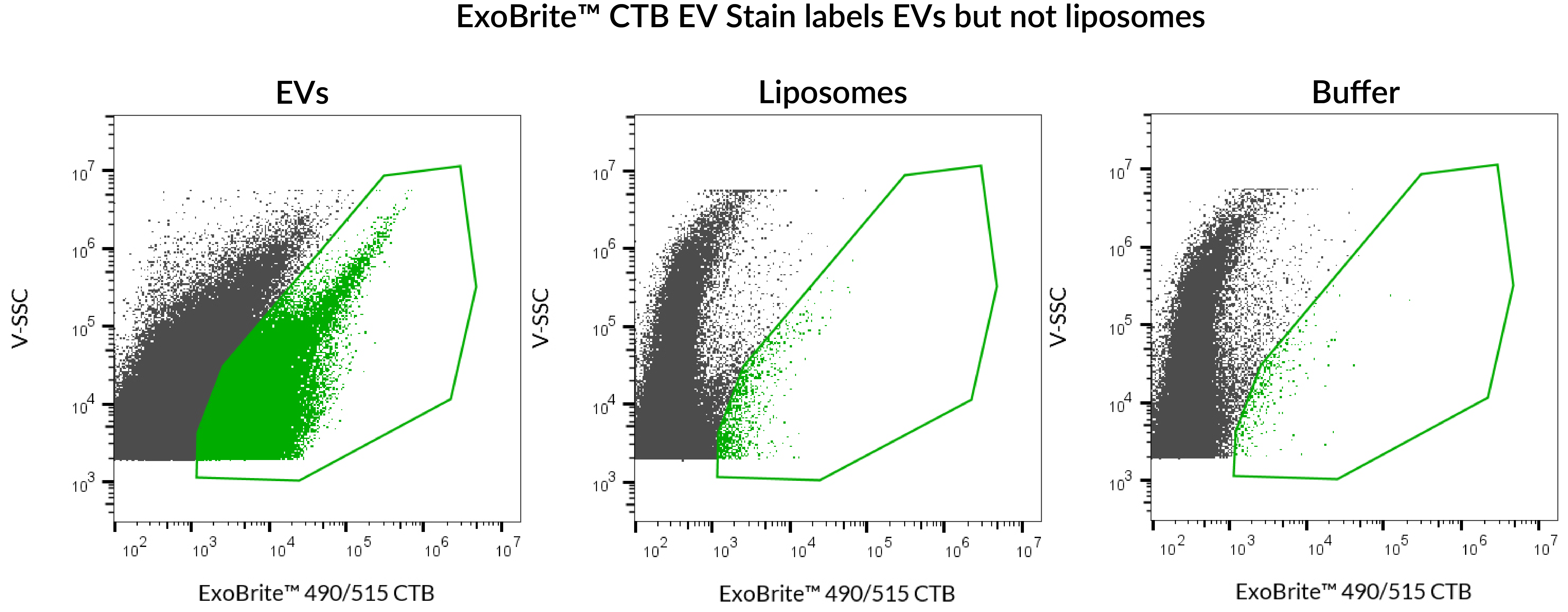 ExoBrite™ 490/515 CTB EV Stain was used to stain either SEC-purified, Jurkat-derived EVs, or artificial liposomes. Robust staining was seen with the EVs, but the liposomes were not stained. The liposomes in this experiment were prepared from Presome® ACD-1, and were verified to be of similar size and concentration to the EVs using the lipophilic dye di-8-ANNEPS (data not shown). Analysis was performed on a CytoFLEX flow cytometer with SSC detected from the 405 nm laser, and ExoBrite™ detected in the FITC channel. Presome® is a registered trademark of NIPPON FINE CHEMICAL CO., LTD.
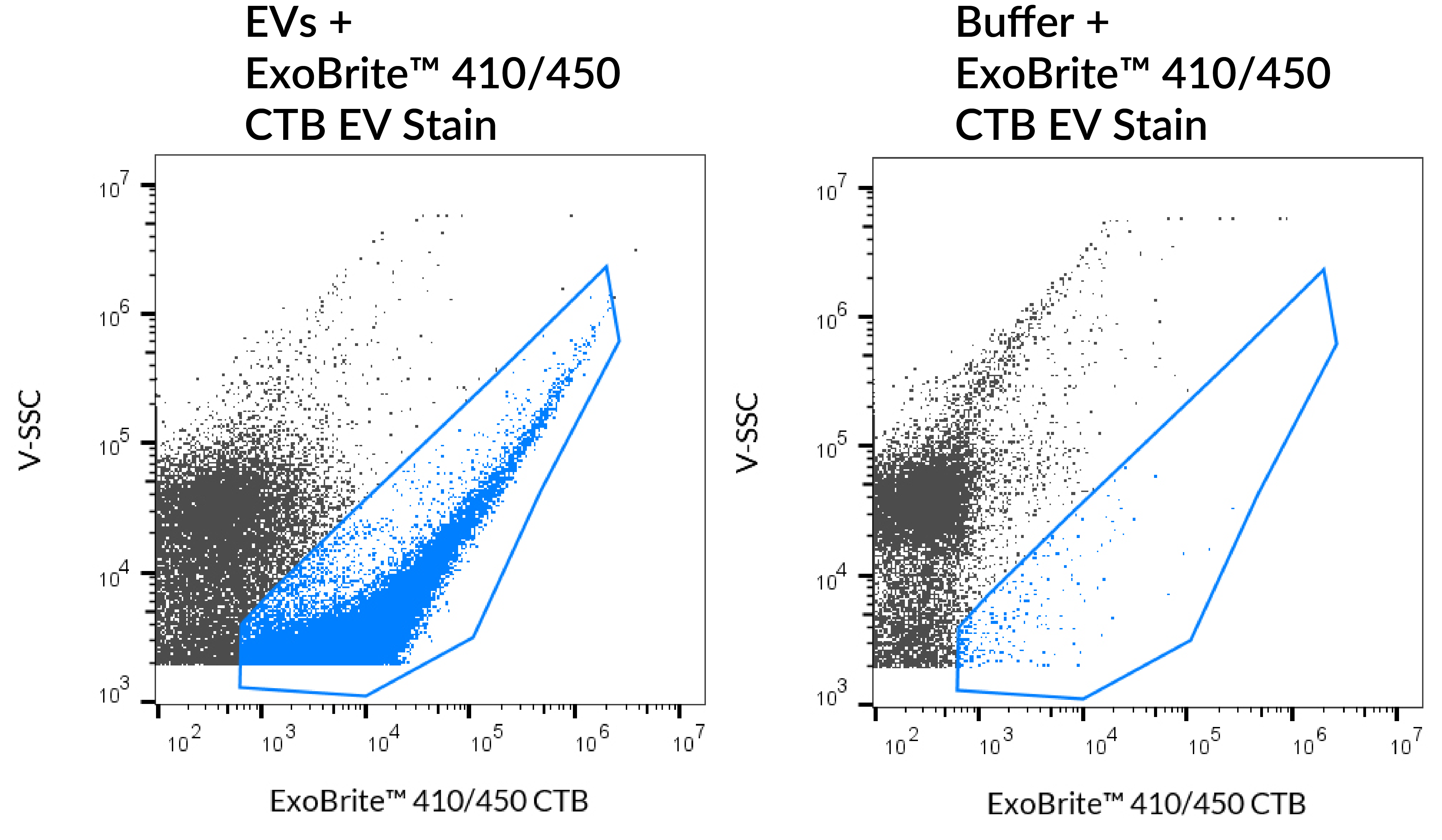 SEC purified MCF-7-derived EVs stained with ExoBrite™ 410/450 CTB EV Stain (left) compared to the same stain in buffer (right). EVs were detected on a CytoFLEX LX flow cytometer in the Pacific Blue channel.
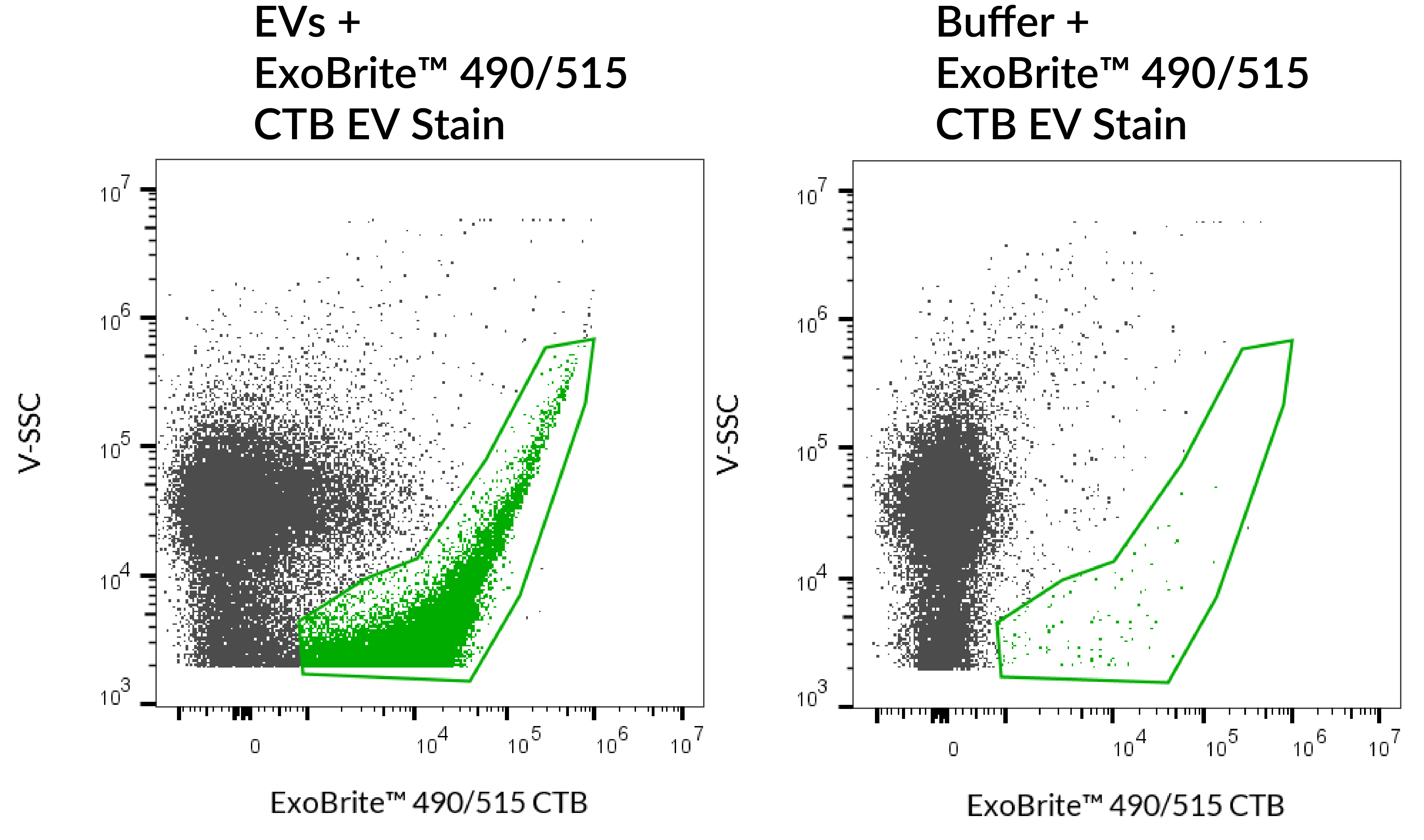 SEC purified MCF-7-derived EVs stained with ExoBrite™ 490/515 CTB EV Stain (left) compared to the same stain in buffer (right). EVs were detected on a CytoFLEX LX flow cytometer in the FITC channel.
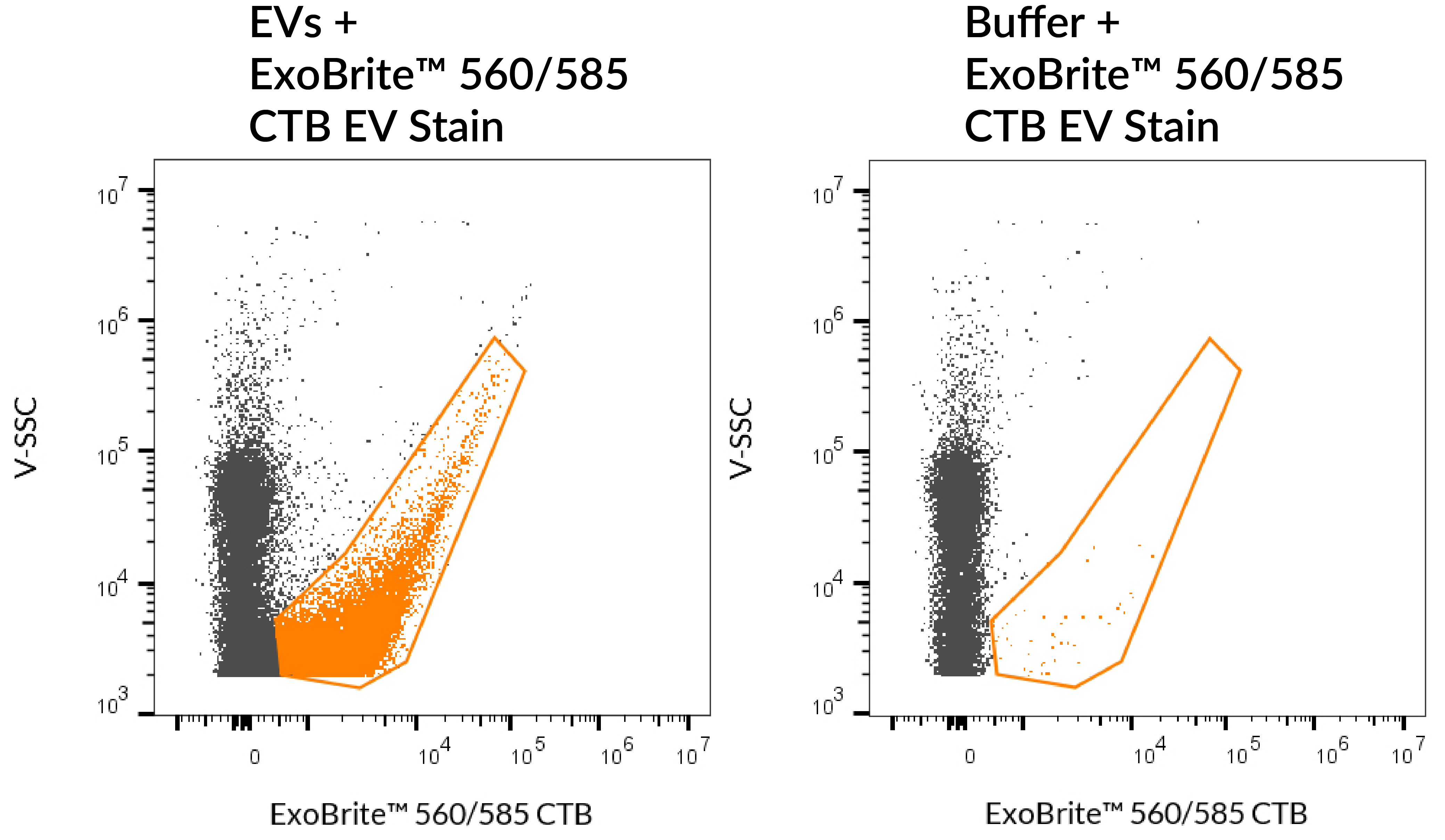 SEC purified MCF-7-derived EVs stained with ExoBrite™ 560/585 CTB EV Stain (left) compared to the same stain in buffer (right). EVs were detected on a CytoFLEX LX flow cytometer in the PE channel.
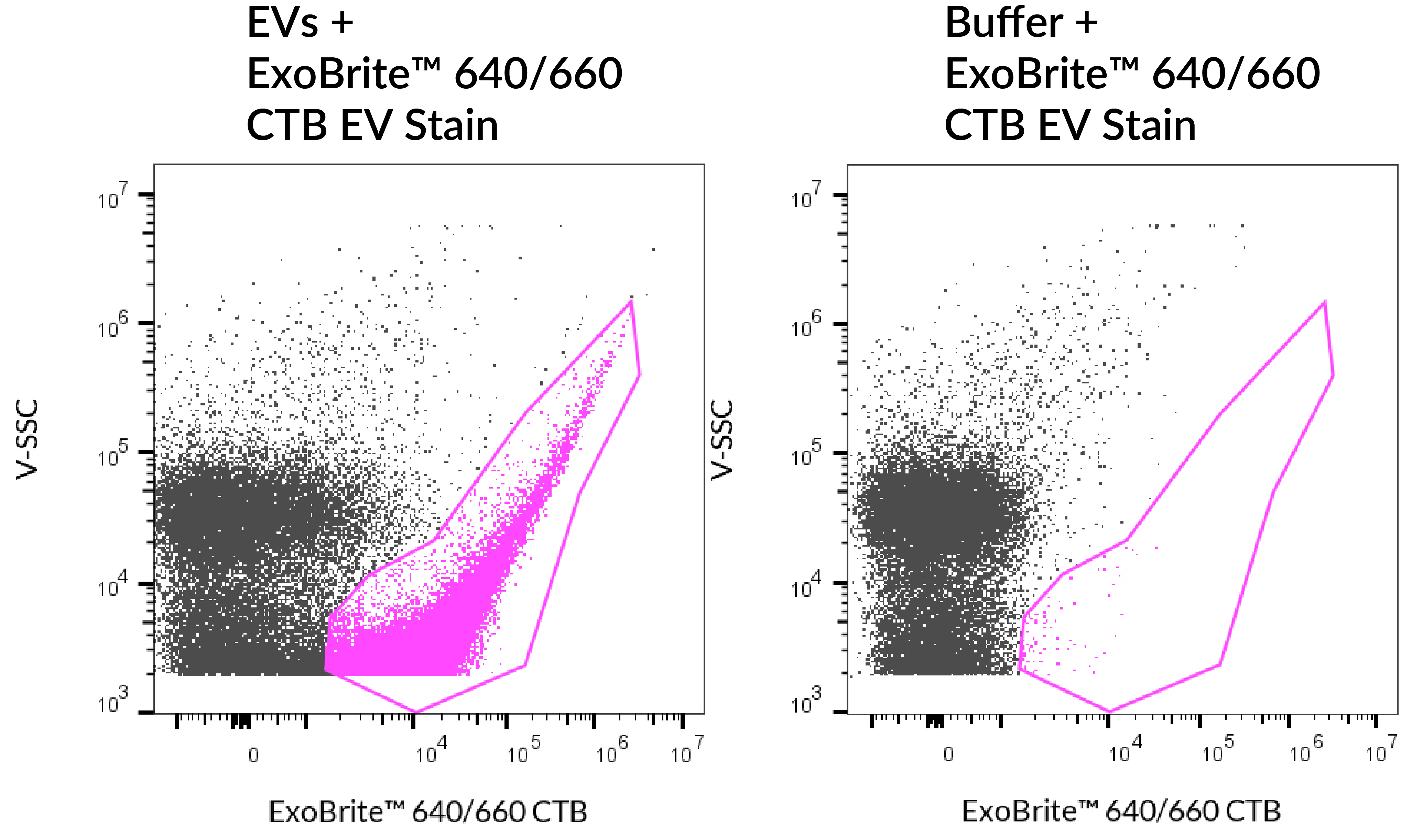 SEC purified MCF-7-derived EVs stained with ExoBrite™ 640/660 CTB EV Stain (left) compared to the same stain in buffer (right). EVs were detected on a CytoFLEX LX flow cytometer in the APC channel.
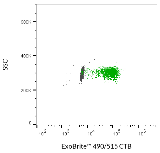 MCF-7-derived EVs were captured onto CD63 beads. The plot shows unstained EVs (gray) or EVs stained with 10X ExoBrite™ 490/515 CTB EV Stain (green). EVs were detected on a CytoFLEX LX flow cytometer in the FITC channel.
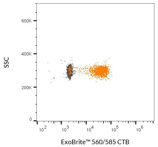 MCF-7-derived EVs were captured onto CD63 beads. The plot shows unstained EVs (gray) or EVs stained with 10X ExoBrite™ 560/585 CTB EV Stain (orange). EVs were detected on a CytoFLEX LX flow cytometer in the PE channel.
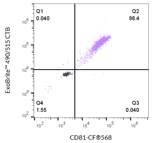 Flow cytometry of CD63 bead-bound EVs derived from MCF-7 cells. Unstained EVs (gray) or co-stained with 10X ExoBrite™ 490/515 CTB and CD81-CF®568 (lavender). EVs were detected on a CytoFLEX LX flow cytometer in the FITC and PE channels.
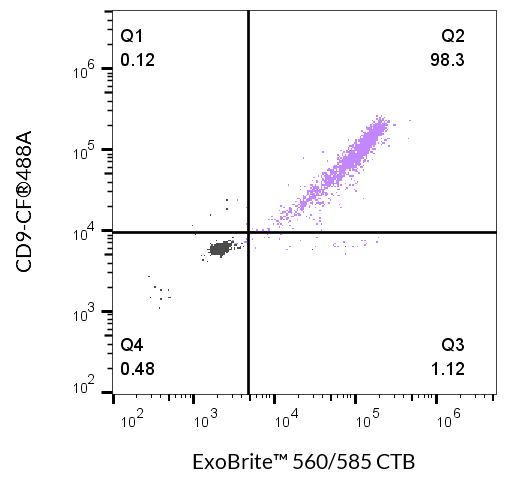 Flow cytometry of CD63 bead-bound EVs derived from MCF-7 cells. Unstained EVs (gray) or co-stained with 10X ExoBrite™ 560/585 CTB and CD9-CF®488A (lavender). EVs were detected on a CytoFLEX LX flow cytometer in the FITC and PE channels.
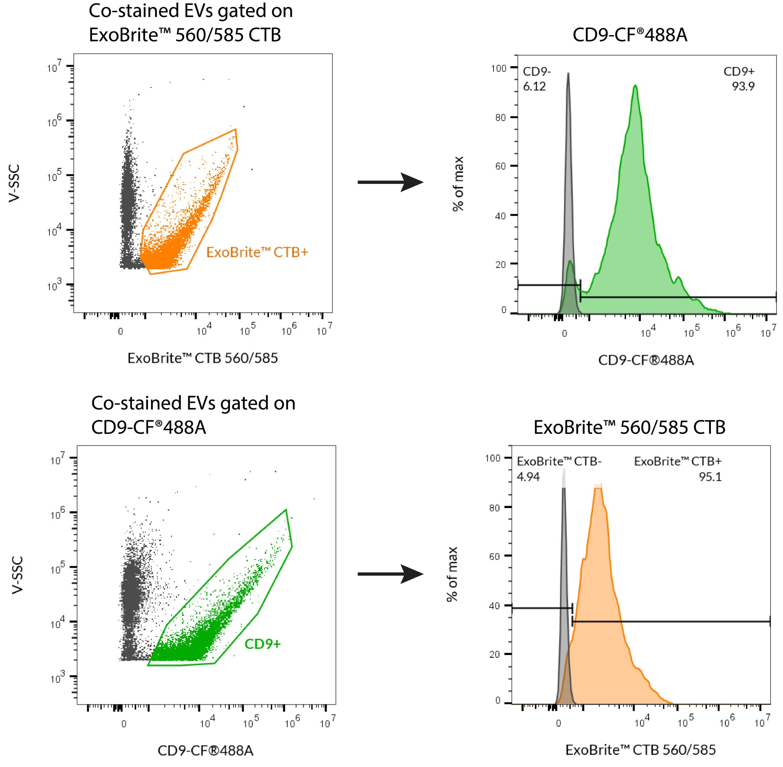 ExoBrite™ CTB EV Stains can be used with antibody co-staining. SEC-purified MCF-7-derived EVs were stained first with 1 ug/mL CD9(H19a)-CF®488A in 100 uL, followed by 10X ExoBrite™ 560/585 CTB EV Stain. EVs were detected on a CytoFLEX LX flow cytometer in the FITC and R-PE channels. Reciprocal gating plots show that when we gated on ExoBrite™ 560/585 CTB-positive particles, ~95% were also positive for CD9 (top row). Likewise, when we gated on CD9-positive particles, ~ 95% were also positive for ExoBrite™ 560/585 CTB (bottom row). Note that MCF-7 cell-derived EVs express high levels of CD9, and CD9 levels differ between cell types.
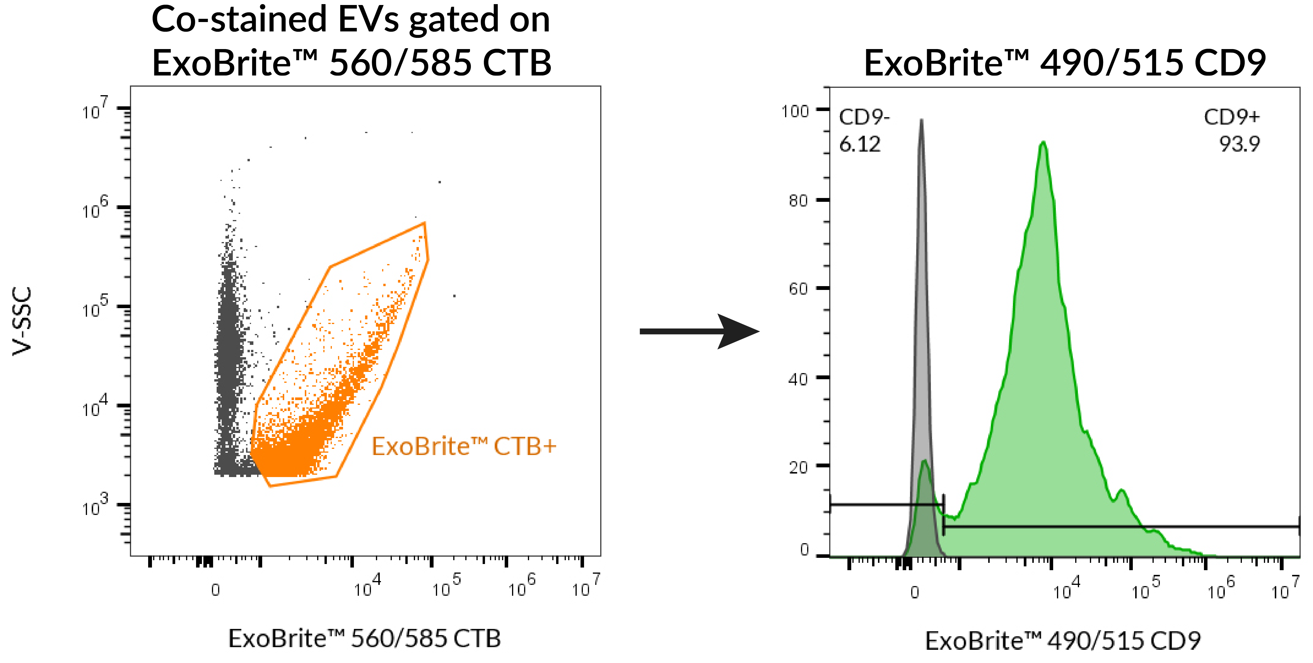 SEC-purified MCF-7-derived EVs were stained first with ExoBrite™ 490/515 CD9 in 100 uL, followed by 10X ExoBrite™ 560/585 CTB EV Stain. EVs were detected on a CytoFLEX LX flow cytometer in the FITC and R-PE channels. When we gated on ExoBrite™ 560/585 CTB-positive particles, ~95% were also positive for CD9.
Product Description
ExoBrite™ CTB EV Staining Kits were designed to overcome some of the challenges of EV detection, particularly in flow cytometry. ExoBrite™ CTB EV Stains bind to molecules in the EV membrane for bright, specific staining, with little to no background.
Features
- Optimally formulated CTB conjugates for staining EVs
- Designed for detection by flow cytometry
- Bright signal and low background
- Stain purified or bead-bound EVs
- Compatible with antibody co-staining
- Available in 4 colors
Kit Components
- ExoBrite™ CTB EV Stain
- ExoBrite™ Reconstitution Solution
- 1 vial of stain makes 100 uL of 500X staining solution
Note: The name of this product has been revised from ExoBrite™ EV Membrane Staining Kits.
ExoBrite™ CTB EV Stains are optimally formulated fluorescent conjugates of cholera toxin subunit B (CTB), which binds to GM1 gangliosides that are commonly found on the surface of mammalian lipid rafts and EVs. The stains were designed to overcome some of the challenges of EV detection, particularly in flow cytometry. Some dyes used to stain EVs can form aggregates of a similar size as exosomes or EVs, thus confounding analysis. ExoBrite™ CTB EV Stains, however, were formulated to show little to no aggregation in flow cytometry, allowing EVs to be identified with bright and specific staining. Unlike hydrophobic membrane dyes, ExoBrite™ CTB EV Stains do not bind non-specifically to polystyrene beads, meaning that they can be used to stain bead-bound EVs.
EVs are often labeled with fluorescent antibodies targeting one or more of the tetraspanin proteins CD9, CD63, and CD81. ExoBrite™ CTB staining can be combined with antibody staining, for multi-parameter analysis.
Less background and better coverage over other EV stains
ExoBrite™ CTB EV Stains were designed to offer exceptional signal:noise and more complete coverage of purified and bead-bound EVs. In the figure below, lipophilic dye DiO and plasma membrane stain CellMask™ demonstrate an unacceptable amount of dye aggregation in gated EVs. Other EV stains such as ExoFlow-ONE™ and ExoGlow™ have less coverage of EVs when compared to ExoBrite™ CTB EV Stains.
ExoBrite™ CTB EV Stains have less background and more complete staining of extracellular vesicles (EVs) than other classic and competitor dyes. EVs were purified from MCF-7 cell supernatant using size exclusion chromatography (SEC). The purified EVs were stained in PBS with the indicated dyes (top row). The stained EV population was gated, with the number showing the percentage of particles falling within the exosome gate. Each dye was also added to filtered PBS (bottom row), to look for dye aggregation and non-specific background. Red arrows indicate these dye aggregates. Lipophilic dyes like DiO show a high number of particles of similar size to EVs, making them unsuited for small particle staining. CellMask™ also shows an unacceptable amount of dye aggregation falling within the EV gate. The ExoFlow-ONE™ and ExoGlow™ dyes form aggregates that can mostly be gated away from the EVs, but they also show less-complete coverage of EVs than ExoBrite™ CTB stains. (Click to enlarge).
Notes:
- ExoBrite™ CTB EV Stains have been found to label EVs derived from several tested cell lines (see Validated EV Sources below), but may not stain EVs from every source.
- In our testing, we have found that ExoBrite™ 490/515 dye may bind to streptavidin coated surfaces or beads if free biotin binding sites are not blocked. We recommend performing a biotin blocking step after binding your biotinylated capture antibody to streptavidin beads or surfaces when using ExoBrite™ 490/515 conjugates. Alternatively, consider using a different ExoBrite™ dye for staining EVs captured on streptavidin beads or surfaces.
ExoBrite™ CTB EV Staining Kits
| Product | Ex/Em | Detection channels | Size | Catalog Number |
|---|---|---|---|---|
| ExoBrite™ 410/450 CTB EV Staining Kit | 416/452 nm | Pacific Blue™ | 100 Labelings | 30111-T |
| 500 Labelings | 30111 | |||
| ExoBrite™ 490/515 CTB EV Staining Kit | 490/516 nm | FITC | 100 Labelings | 30112-T |
| 500 Labelings | 30112 | |||
| ExoBrite™ 560/585 CTB EV Staining Kit | 562/584 nm | PE, Cy®3 | 100 Labelings | 30113-T |
| 500 Labelings | 30113 | |||
| ExoBrite™ 640/660 CTB EV Staining Kit | 642/663 nm | APC | 100 Labelings | 30114-T |
| 500 Labelings | 30114 |
Validated EV Sources for ExoBrite™ EV Surface Stains
| EV Source | ExoBrite™ True EV Membrane Stains | ExoBrite™ CTB Stains | ExoBrite™ WGA Stains | ExoBrite™ Annexin Stains |
|---|---|---|---|---|
| A549 cells | Yes | Yes | Yes | Yes |
| CHO cells | Yes | No | Yes | Yes |
| hASC (human adipose stem cells) | ND | No1 | ND | ND |
| HEK293 cells | Yes | Yes1 | Yes | Yes |
| HeLa cells | Yes | No | Yes | Yes |
| HUVEC (human umbilical vein endothelial cells) | ND | No1 | ND | ND |
| J774 cells | Yes | Yes | Yes | Yes |
| Jurkat cells | Yes | Yes | Yes | Yes |
| MCF-7 cells | Yes | Yes | Yes | Yes |
| Plasma | Yes | No | ND | Yes |
| Raji cells | ND | Yes | Yes | Yes |
| RAW 264.7 cells | Yes | Yes | Yes | Yes |
| Serum | Yes | No | ND | Yes |
| Skeletal myoblasts | ND | Yes1 | ND | ND |
| THP-1 cells | Yes | ND | ND | ND |
| U2OS cells | Yes | No | Yes | Yes |
| U937 cells | Yes | No | Yes | Yes |
| NIH3T3 cells | Yes | Yes | Yes | Yes |
| HepG2 cells | Yes | No | Yes | Yes |
| Yeast (S. cerevisiae) | Yes | No | Yes | Yes |
Value of “Yes” or “No” indicates coverage of EVs based on Biotium’s internal data or customer-reported data. Value of “ND” indicates no data.
ExoBrite™ EV Stains Comparison Guide
| ExoBrite™ EV Surface Stain | Pros | Cons |
|---|---|---|
| ExoBrite™ True EV Membrane Stains | • Near-complete staining of EVs in a sample • Broad compatibility with different EV sources • Validated for flow and fNTA | • Can't be used to stain bead-bound EVs • May have more aggregation than CTB & Annexin |
| ExoBrite™ Annexin EV Staining Kits | • Broad compatibility with different EV sources • Validated for flow and fNTA • Low background aggregates | • May not stain every EV in a sample • Doesn't work well on bead-bound EVs |
| ExoBrite™ WGA EV Staining Kits | • Broad compatibility with different EV sources • Can be used with bead-bound EVs | • May not stain every EV in a sample • Doesn't work well for fNTA |
| ExoBrite™ CTB EV Staining Kits | • Validated for flow and fNTA • Extremely low background • Can be used with bead-bound EVs | • May not stain every EV in a sample • Does not stain EVs from every source |
| ExoBrite™ Antibodies | • Highly specific for human tetraspanins CD9, CD63, CD81, and other EV markers • Validated for EV flow • Broad compatibility for different EV sources • Can be used with bead-bound EVs • Can be used for WB | • Depends on the expression level of the target protein on the EVs |
| ExoBrite™ EV Stain Enhancer | • Improves signal-to-noise by reducing or eliminating aggregates of certain EV stains • Validated with several different lectins and Annexin V • Does not interfere with antibody staining of EVs • Easy to use, just add directly to the staining reaction | • Not recommended for use with lipophilic EV stains |
Biotium also offers other validated ExoBrite™ reagents for flow cytometry, western blotting, or super-resolution imaging.
Learn about Biotium's new ExoBrite™ True EV Membrane Stains. These genuine lipophilic membrane dyes are designed for superior pan-EV labeling over other membrane dyes including PKH, DiO, DiI, and DiD. Biotium also offers ExoBrite™ WGA EV Stains (wheat germ agglutinin) and ExoBrite™ Annexin EV Stains optimized for bright and sensitive staining of EVs. The ExoBrite™ EV Surface Stain Sampler Kit contains each of Biotium’s ExoBrite™ EV Surface Stains (CTB, WGA, and Annexin V) for assessing which stain offers the best coverage for the EV samples of interest. Biotium also offers ExoBrite™ Antibody Conjugates for optimal detection of CD9, CD63, and CD81 EV markers by flow cytometry and western blotting. For super-resolution imaging by STORM, learn about our ExoBrite™ STORM CTB EV Staining Kits available in four CF® Dyes validated for STORM.
Technology

Highlighted Citations

Tech Tips

The Full Spectrum Blog

