
PPAR alpha antibody


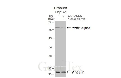
Non-transfected (–) and transfected (+) unboiled HepG2 whole cell extracts (30 μg) were separated by 10% SDS-PAGE, and the membrane was blotted with PPAR alpha antibody (GTX101098) diluted at 1:1000. The HRP-conjugated anti-rabbit IgG antibody (GTX213110-01) was used to detect the primary antibody.
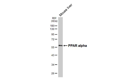
Mouse tissue extract (50 μg) was separated by 10% SDS-PAGE, and the membrane was blotted with PPAR alpha antibody (GTX101098) diluted at 1:1000. The HRP-conjugated anti-rabbit IgG antibody (GTX213110-01) was used to detect the primary antibody.
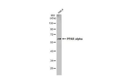
Whole cell extract (30 μg) was separated by 10% SDS-PAGE, and the membrane was blotted with PPAR alpha antibody (GTX101098) diluted at 1:500. The HRP-conjugated anti-rabbit IgG antibody (GTX213110-01) was used to detect the primary antibody.
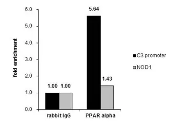
ChIP experiment and primer designs are based on J Biol Chem. 2013 Jan 18;288(3):1726-38.
Cross-linked ChIP was performed with HepG2 chromatin extract and 5 μg of either control rabbit IgG or anti-PPAR alpha antibody. The precipitated DNA was detected by PCR with primer set targeting to C3 promotor or NOD1 gene.
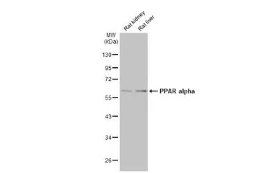
Various tissue extracts (50 μg) were separated by 10% SDS-PAGE, and the membrane was blotted with PPAR alpha antibody (GTX101098) diluted at 1:2000. The HRP-conjugated anti-rabbit IgG antibody (GTX213110-01) was used to detect the primary antibody.
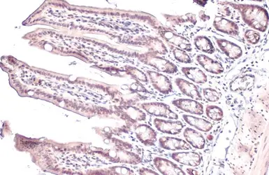
PPAR alpha antibody detects PPAR alpha protein at nucleus by immunohistochemical analysis.Sample: Paraffin-embedded mouse intestine.PPAR alpha stained by PPAR alpha antibody (GTX101098) diluted at 1:165.Antigen Retrieval: Citrate buffer, pH 6.0, 15 min
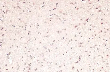
PPAR alpha antibody detects PPAR alpha protein at nucleus by immunohistochemical analysis.Sample: Paraffin-embedded rat brain.PPAR alpha stained by PPAR alpha antibody (GTX101098) diluted at 1:165.Antigen Retrieval: Citrate buffer, pH 6.0, 15 min
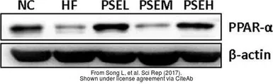
The data was published in the 2017 in Sci Rep. PMID: 28169366
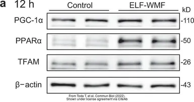
The data was published in the 2022 in Commun Biol. PMID: 35552531