

Rabbit Anti-HIF1A Polyclonal Antibody

Referência AP4879d-ev
Tamanho : 80ul
Marca : Abcepta
HIF1A Antibody
Affinity Purified Rabbit Polyclonal Antibody (Pab)
-
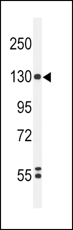
Western blot analysis of HIF1A Antibody (Cat. #AP4879d) in K562 cell line lysates (35ug/lane). HIF1A (arrow) was detected using the purified Pab.
-
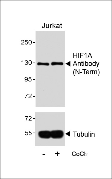
Western blot analysis of lysates from Jurkat cell line, untreated or treated with CoCl2, 0. 1mM, using 157999101(Cat. #AP4879d)(upper) or Tubulin (lower).
-
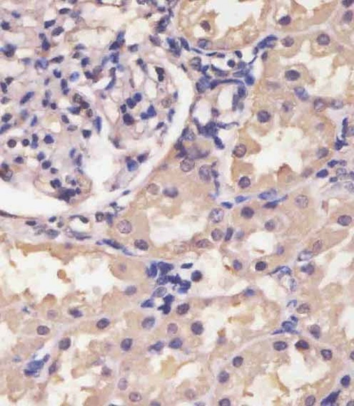
AP4879D staining HIF1A in human kidney tissue sections by Immunohistochemistry (IHC-P - paraformaldehyde-fixed, paraffin-embedded sections). Tissue was fixed with formaldehyde and blocked with 3% BSA for 0. 5 hour at room temperature; antigen retrieval was by heat mediation with a citrate buffer (pH6). Samples were incubated with primary antibody (1/25) for 1 hours at 37°C. A undiluted biotinylated goat polyvalent antibody was used as the secondary antibody.
-
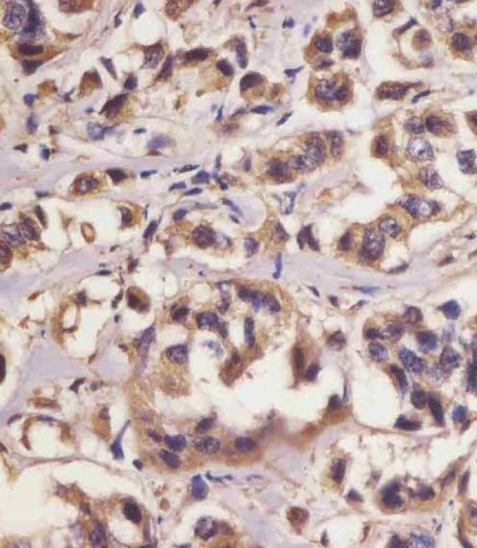
AP4879D staining HIF1A in human breast carcinoma sections by Immunohistochemistry (IHC-P - paraformaldehyde-fixed, paraffin-embedded sections). Tissue was fixed with formaldehyde and blocked with 3% BSA for 0. 5 hour at room temperature; antigen retrieval was by heat mediation with a citrate buffer (pH6). Samples were incubated with primary antibody (1/25) for 1 hours at 37°C. A undiluted biotinylated goat polyvalent antibody was used as the secondary antibody.
-
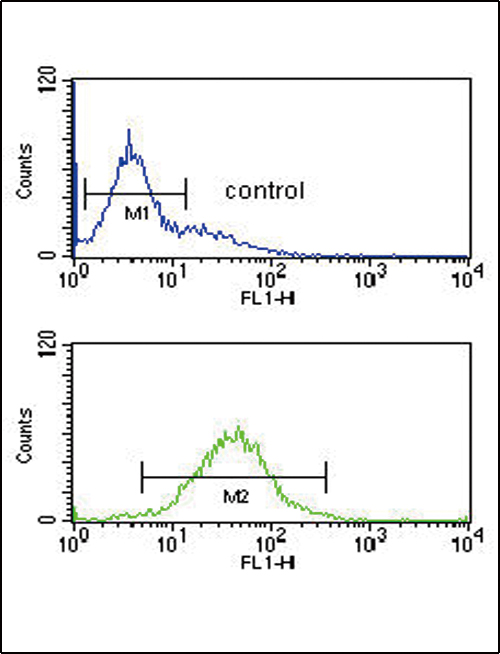
HIF1A Antibody (Cat. #AP4879d) flow cytometric analysis of K562 cells (bottom histogram) compared to a negative control cell (top histogram).FITC-conjugated goat-anti-rabbit secondary antibodies were used for the analysis.
Western blot analysis of HIF1A Antibody (Cat. #AP4879d) in K562 cell line lysates (35ug/lane). HIF1A (arrow) was detected using the purified Pab.
Application 
| WB, FC, IHC-P, E |
|---|---|
| Primary Accession | Q16665 |
| Reactivity | Human |
| Host | Rabbit |
| Clonality | Polyclonal |
| Isotype | Rabbit IgG |
| Calculated MW | 92670 Da |
| Gene ID | 3091 |
|---|---|
| Other Names | Hypoxia-inducible factor 1-alpha, HIF-1-alpha, HIF1-alpha, ARNT-interacting protein, Basic-helix-loop-helix-PAS protein MOP1, Class E basic helix-loop-helix protein 78, bHLHe78, Member of PAS protein 1, PAS domain-containing protein 8, HIF1A, BHLHE78, MOP1, PASD8 |
| Target/Specificity | This HIF1A antibody is generated from rabbits immunized with HIF1A recombinant protein. |
| Dilution | WB~~1:1000 FC~~1:10~50 IHC-P~~1:25 E~~Use at an assay dependent concentration. |
| Format | Purified polyclonal antibody supplied in PBS with 0.09% (W/V) sodium azide. This antibody is purified through a protein A column, followed by peptide affinity purification. |
| Storage | Maintain refrigerated at 2-8°C for up to 2 weeks. For long term storage store at -20°C in small aliquots to prevent freeze-thaw cycles. |
| Precautions | HIF1A Antibody is for research use only and not for use in diagnostic or therapeutic procedures. |
For Research Use Only. Not For Use In Diagnostic Procedures.
| Name | HIF1A {ECO:0000303|PubMed:7539918, ECO:0000312|HGNC:HGNC:4910} |
|---|---|
| Function | Functions as a master transcriptional regulator of the adaptive response to hypoxia (PubMed:11292861, PubMed:11566883, PubMed:15465032, PubMed:16973622, PubMed:17610843, PubMed:18658046, PubMed:20624928, PubMed:22009797, PubMed:30125331, PubMed:9887100). Under hypoxic conditions, activates the transcription of over 40 genes, including erythropoietin, glucose transporters, glycolytic enzymes, vascular endothelial growth factor, HILPDA, and other genes whose protein products increase oxygen delivery or facilitate metabolic adaptation to hypoxia (PubMed:11292861, PubMed:11566883, PubMed:15465032, PubMed:16973622, PubMed:17610843, PubMed:20624928, PubMed:22009797, PubMed:30125331, PubMed:9887100). Plays an essential role in embryonic vascularization, tumor angiogenesis and pathophysiology of ischemic disease (PubMed:22009797). Heterodimerizes with ARNT; heterodimer binds to core DNA sequence 5'-TACGTG-3' within the hypoxia response element (HRE) of target gene promoters (By similarity). Activation requires recruitment of transcriptional coactivators such as CREBBP and EP300 (PubMed:16543236, PubMed:9887100). Activity is enhanced by interaction with NCOA1 and/or NCOA2 (PubMed:10594042). Interaction with redox regulatory protein APEX1 seems to activate CTAD and potentiates activation by NCOA1 and CREBBP (PubMed:10202154, PubMed:10594042). Involved in the axonal distribution and transport of mitochondria in neurons during hypoxia (PubMed:19528298). |
| Cellular Location | Cytoplasm. Nucleus. Nucleus speckle {ECO:0000250|UniProtKB:Q61221}. Note=Colocalizes with HIF3A in the nucleus and speckles (By similarity). Cytoplasmic in normoxia, nuclear translocation in response to hypoxia (PubMed:9822602) {ECO:0000250|UniProtKB:Q61221, ECO:0000269|PubMed:9822602} |
| Tissue Location | Expressed in most tissues with highest levels in kidney and heart. Overexpressed in the majority of common human cancers and their metastases, due to the presence of intratumoral hypoxia and as a result of mutations in genes encoding oncoproteins and tumor suppressors. A higher level expression seen in pituitary tumors as compared to the pituitary gland. |
- Primary Antibodies
- Cancer
- Cardiovascular
- Metabolism