


Vimentin antibody




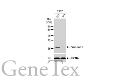
Wild-type (WT) and Vimentin knockout (KO) 293T cell extracts (30 μg) were separated by 10% SDS-PAGE, and the membrane was blotted with Vimentin antibody (GTX100619) diluted at 1:50000. The HRP-conjugated anti-rabbit IgG antibody (GTX213110-01) was used to detect the primary antibody.
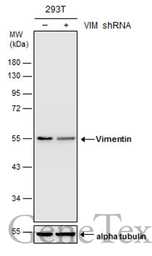
Non-transfected (–) and transfected (+) 293T whole cell extracts (30 μg) were separated by 10% SDS-PAGE, and the membrane was blotted with Vimentin antibody (GTX100619) diluted at 1:20000.
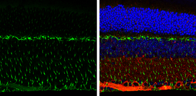
Vimentin antibody detects Vimentin protein expression by immunohistochemical analysis.
Sample:Paraffin-Embedded adult mouse retina.
Green: Vimentin protein stained by Vimentin antibody (GTX100619) diluted at 1:250.
Red: beta Tubulin 3/ TUJ1, stained by beta Tubulin 3/ TUJ1 antibody [GT11710] (GTX631836) diluted at 1:500.
Blue: Fluoroshield with DAPI (GTX30920).
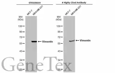
Various whole cell extracts (30 μg) were separated by 10% SDS-PAGE, and the membranes were blotted with Vimentin antibody (GTX100619) diluted at 1:5000 and competitor's antibody diluted at 1:5000. The HRP-conjugated anti-rabbit IgG antibody (GTX213110-01) was used to detect the primary antibody.
*The competitor is not affiliated with GeneTex and does not endorse this product.
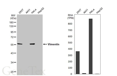
Various whole cell extracts (30 μg) were separated by 10% SDS-PAGE, and the membrane was blotted with Vimentin antibody (GTX100619) diluted at 1:50000. The HRP-conjugated anti-rabbit IgG antibody (GTX213110-01) was used to detect the primary antibody. Corresponding RNA expression data for the same cell lines are based on Human Protein Atlas program.
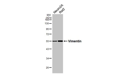
Various whole cell extracts (30 μg) were separated by 10% SDS-PAGE, and the membrane was blotted with Vimentin antibody (GTX100619) diluted at 1:2000. The HRP-conjugated anti-rabbit IgG antibody (GTX213110-01) was used to detect the primary antibody.
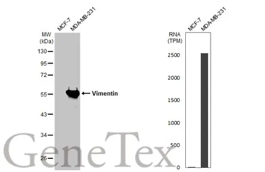
Various whole cell extracts (30 μg) were separated by 10% SDS-PAGE, and the membrane was blotted with Vimentin antibody (GTX100619) diluted at 1:5000. The HRP-conjugated anti-rabbit IgG antibody (GTX213110-01) was used to detect the primary antibody. Corresponding RNA expression data for the same cell lines are based on Human Protein Atlas program.
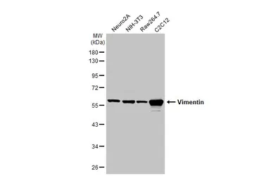
Various whole cell extracts (30 μg) were separated by 10% SDS-PAGE, and the membrane was blotted with Vimentin antibody (GTX100619) diluted at 1:5000. The HRP-conjugated anti-rabbit IgG antibody (GTX213110-01) was used to detect the primary antibody.
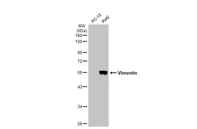
Various whole cell extracts (30 μg) were separated by 10% SDS-PAGE, and the membrane was blotted with Vimentin antibody (GTX100619) diluted at 1:10000. The HRP-conjugated anti-rabbit IgG antibody (GTX213110-01) was used to detect the primary antibody.
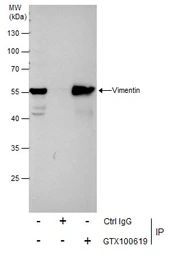
Immunoprecipitation of Vimentin protein from HeLa whole cell extracts using 5 μg of Vimentin antibody (GTX100619).
Western blot analysis was performed using Vimentin antibody (GTX100619).
EasyBlot anti-Rabbit IgG (GTX221666-01) was used as a secondary reagent.
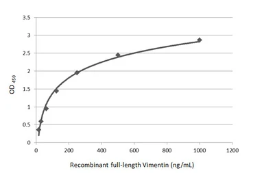
Sandwich ELISA detection of recombinant full-length Vimentin protein using GTX629744 as capture antibody at concentration of 5 μg/mL and GTX100619 as detection antibody at concentration of 1 μg/mL. Rabbit IgG antibody (HRP) (GTX213110-01) was diluted at 1:10000 and used to detect the primary antibody.
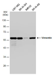
Various whole cell extracts (30 μg) were separated by 10% SDS-PAGE, and the membrane was blotted with Vimentin antibody (GTX100619) diluted at 1:50000. The HRP-conjugated anti-rabbit IgG antibody (GTX213110-01) was used to detect the primary antibody.
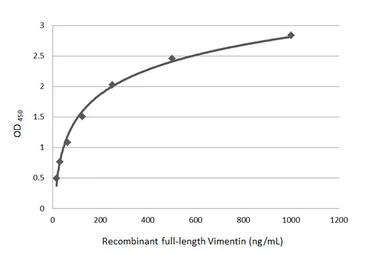
Sandwich ELISA detection of recombinant full-length Vimentin protein using GTX629743 as capture antibody at concentration of 5 μg/mL and GTX100619 as detection antibody at concentration of 1 μg/mL. Rabbit IgG antibody (HRP) (GTX213110-01) was diluted at 1:10000 and used to detect the primary antibody.
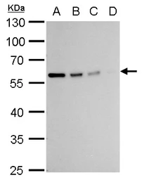
Vimentin antibody detects Vimentin protein by western blot analysis.
A. 20 μg 293T whole cell lysate/extract
B. 10 μg 293T whole cell lysate/extract
C. 5 μg 293T whole cell lysate/extract
D. 1 μg 293T whole cell lysate/extract
10% SDS-PAGE
Vimentin antibody (GTX100619) dilution: 1:10000
The HRP-conjugated anti-rabbit IgG antibody (GTX213110-01) was used to detect the primary antibody.
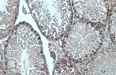
Vimentin antibody detects Vimentin protein at cytoplasm by immunohistochemical analysis.Sample: Paraffin-embedded mouse testis.Vimentin stained by Vimentin antibody (GTX100619) diluted at 1:500.Antigen Retrieval: Citrate buffer, pH 6.0, 15
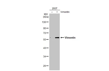
Non-transfected (–) and transfected (+) 293T whole cell extracts (10 μg) were separated by 10% SDS-PAGE, and the membrane was blotted with Vimentin antibody (GTX100619) diluted at 1:10000. The HRP-conjugated anti-rabbit IgG antibody (GTX213110-01) was used to detect the primary antibody.
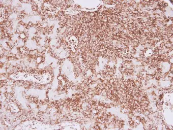
Vimentin antibody detects Vimentin protein at cytoplasm in human lung adenocarcinoma by immunohistochemical analysis.
Sample: Paraffin-embedded human lung adenocarcinoma.
Vimentin antibody (GTX100619) diluted at 1:500.
Antigen Retrieval: Trilogy™ (EDTA based, pH 8.0) buffer, 15min
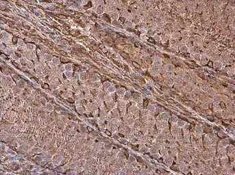
Vimentin antibody detects Vimentin protein at cell membrane and cytoplasm in rat testis by immunohistochemical analysis.
Sample: Paraffin-embedded rat testis.
Vimentin antibody (GTX100619) diluted at 1:500.
Antigen Retrieval: Citrate buffer, pH 6.0, 15 min
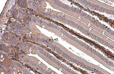
Vimentin antibody detects Vimentin protein at cytoplasm by immunohistochemical analysis.Sample: Paraffin-embedded mouse intestine.Vimentin stained by Vimentin antibody (GTX100619) diluted at 1:500.Antigen Retrieval: Citrate buffer, pH 6.0, 15 min
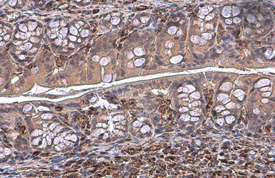
Vimentin antibody detects Vimentin protein at cytoplasm by immunohistochemical analysis.Sample: Paraffin-embedded mouse colon.Vimentin stained by Vimentin antibody (GTX100619) diluted at 1:500.Antigen Retrieval: Citrate buffer, pH 6.0, 15 min
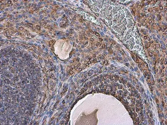
Vimentin antibody detects Vimentin protein at cell membrane and cytoplasm in rat ovary by immunohistochemical analysis.
Sample: Paraffin-embedded rat ovary.
Vimentin antibody (GTX100619) diluted at 1:500.
Antigen Retrieval: Citrate buffer, pH 6.0, 15 min
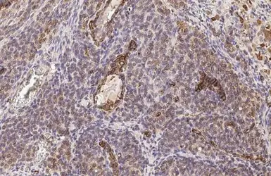
Vimentin antibody detects Vimentin protein at cell membrane and cytoplasm by immunohistochemical analysis.Sample: Paraffin-embedded rat ovary.Vimentin stained by Vimentin antibody (GTX100619) diluted at 1:500.Antigen Retrieval: Citrate buffer, pH 6.0, 15 min
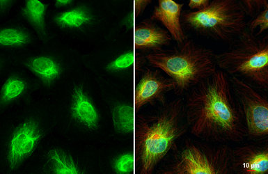
Vimentin antibody detects Vimentin protein at cytoskeleton and nucleus by immunofluorescent analysis.Sample: HeLa cells were fixed in 4% paraformaldehyde at RT for 15 min.Green: Vimentin stained by Vimentin antibody (GTX100619) diluted at 1:500.Red: alpha Tubulin, a cytoskeleton marker, stained by alpha Tubulin antibody [GT114] (GTX628802) diluted at 1:1000.Blue: Fluoroshield with DAPI (GTX30920).
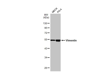
Various whole cell extracts (30 μg) were separated by 10% SDS-PAGE, and the membrane was blotted with Vimentin antibody (GTX100619) diluted at 1:20000. The HRP-conjugated anti-rabbit IgG antibody (GTX213110-01) was used to detect the primary antibody.
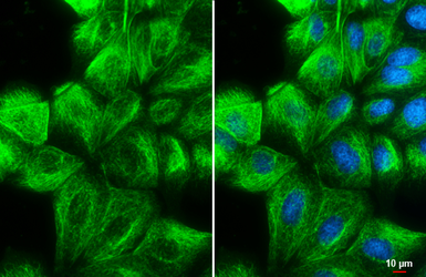
Vimentin antibody detects Vimentin protein at cytoskeleton by immunofluorescent analysis.Sample: MDCK cells were fixed in 4% paraformaldehyde at RT for 15 min.Green: Vimentin stained by Vimentin antibody (GTX100619) diluted at 1:500.Blue: Hoechst 33342 staining.Scale bar= 10 μm.
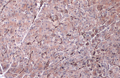
Vimentin antibody detects Vimentin protein by immunohistochemical analysis.Sample: Paraffin-embedded dog pancreas.Vimentin stained by Vimentin antibody (GTX100619) diluted at 1:500.Antigen Retrieval: Citrate buffer, pH 6.0, 15 min
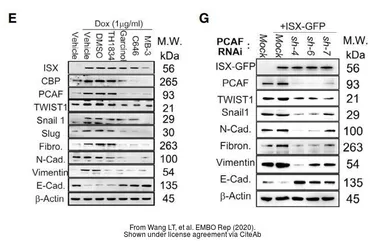
The data was published in the journal EMBO Rep in 2020. PMID: 31908141
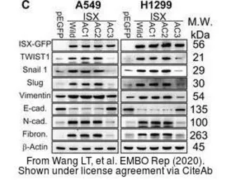
The data was published in the journal EMBO Rep in 2020. PMID: 31908141
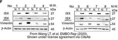
The data was published in the journal EMBO Rep in 2020. PMID: 31908141
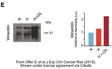
The data was published in the journal J Exp Clin Cancer Res in 2019. PMID: 31174567
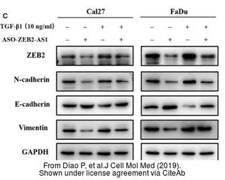
The data was published in the journal J Cell Mol Med in 2019. PMID: 30950191

The data was published in the journal J Biomed Sci in 2013. PMID: 24359494
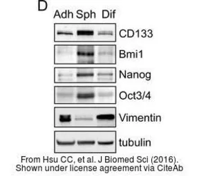
The data was published in the journal J Biomed Sci in 2016.PMID: 27863490
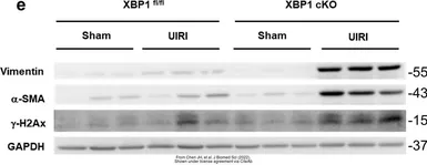
The data was published in the 2022 in J Biomed Sci. PMID: 35765067
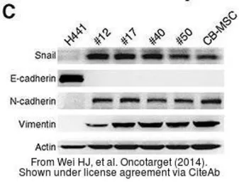
The data was published in the journal Oncotarget in 2014. PMID: 25237908
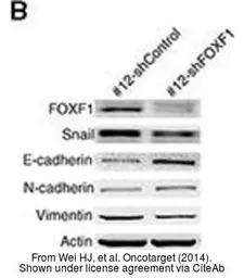
The data was published in the journal Oncotarget in 2014. PMID: 25237908
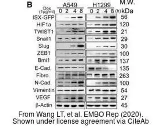
The data was published in the journal EMBO Rep in 2020. PMID: 31908141
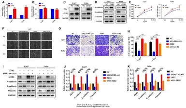
The data was published in the journal J Cell Mol Med in 2019. PMID: 30950191
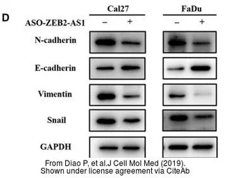
The data was published in the journal J Cell Mol Med in 2019. PMID: 30950191
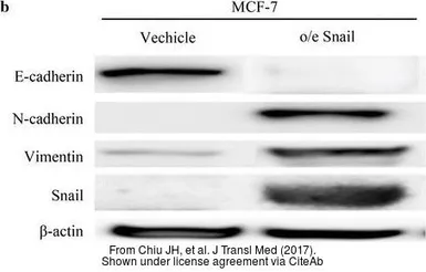
The data was published in the journal J Transl Med in 2017. PMID: 28472954
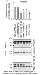
The data was published in the journal Nat Commun in 2015. PMID: 26245647
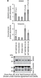
The data was published in the journal Nat Commun in 2015. PMID: 26245647
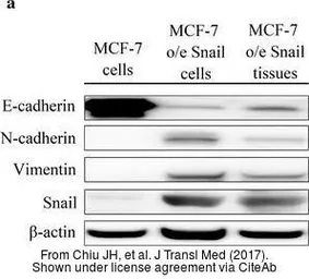
The data was published in the journal J Transl Med in 2017. PMID: 28472954
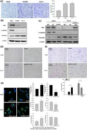
The data was published in the journal Sci Rep in 2017. PMID: 28195146
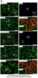
The data was published in the journal PLoS One in 2014. PMID: 25397403
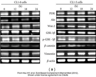
The data was published in the journal Evid Based Complement Alternat Med in 2012.PMID: 23320038
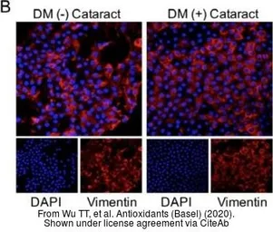
The data was published in the journal Antioxidants (Basel) in 2020.PMID: 32218152
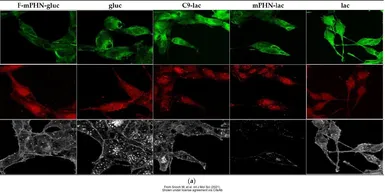
The data was published in the 2021 in Int J Mol Sci. PMID: 34281292