

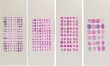
Paraffin tissue arrays, commonly referred to as paraffin tissue microarrays (TMA), are composite paraffin blocks containing multiple small tissue cores sampled from paraffin-embedded donor tissues, typically formalin-fixed, paraffin-embedded (FFPE) specimens, and arranged at defined coordinates. Sections from the recipient block enable multiple specimens to be analyzed in parallel under comparable experimental conditions, supporting high-throughput histological and molecular studies while conserving valuable tissue resources.
Arrays can be constructed from tissues of the same species, allowing comparisons among organs, pathological conditions, experimental groups, or individual specimens. Multi-species tissue arrays can incorporate tissues from different animal species and are particularly relevant to comparative pathology and veterinary research, including comparative assessment of tissue or tumor biomarker expression. Veterinary studies have demonstrated the use of TMAs for simultaneous immunohistochemical analysis of multiple animal tissues and neoplasms.
Key Features
-
Multiple Paraffin-Embedded Tissue Cores : Organized within a single recipient block.
-
Same-Species or Multi-Species Configurations : Supports comparative research across tissues, species, pathological conditions, or experimental groups.
-
Parallel Analysis : Enables analysis of numerous specimens on the same slide using standardized assay conditions.
-
Tissue and Reagent Conservation : Reduces consumption of valuable donor tissue, slides, and reagents.
-
Broad Analytical Compatibility : Compatible with histology, immunohistochemistry (IHC), fluorescence in situ hybridization (FISH), and RNA in situ hybridization (RNA-ISH), as well as other in situ molecular assays.
Usual Research Applications
-
Biomarker validation and protein-expression profiling.
-
Molecular pathology and comparative pathology.
-
Disease-model research and tumor characterization.
-
High-throughput tissue analysis across multiple specimens or experimental groups.
-
Systematic image acquisition, quantitative assessment, and comparative analysis using defined tissue coordinates.