

CellSurface FLOW Staining Kit

Cat# MBS668926-5x1
Size : 5x1Kit
Brand : MyBiosource
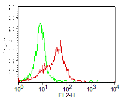
Cell Surface Staining of PBMC. 0.5 ug antibody was used. Green: Isotype control (mouse IgG1 PE). Red: human TLR2 PE. Cell surface staining kit was used.
Cellsurface Staining Kit
For Research Use Only. Not for use in diagnostic procedures. Catalog #: MBS668926The Cell Surface FLOW Staining Kit allows the detection of antibodies on the cellsurface proteins in Flowcytometry analysis. This kit is specially formulated to reduce non-specific staining of fluorochrome-labelled antibodies and to increase fluorescence… More details
Product Overview
Online Datasheet in PDFProduct Name
CellSurface, Staining Kit
Full Product Name
CellSurface FLOW Staining Kit
Product Synonym Names
CellSurface Staining FLOW Kits and Reagents; Kits and Reagents
Preparation and Storage
Store at 2-8 degree C
Staining Buffer
1X - 4 X 60ml
Fixation Buffer
10% - 15ml
Product Categories/Family for CellSurface staining kit
Kits and Reagents; Flowcytometry staining Kits
Applications Tested/Suitable for CellSurface staining kit
Flow Cytometry (FC/FACS)
Application Notes for CellSurface staining kit
Protocol: 1. Determine number of cells required for staining. Each sample contains 0.5 -1 x 10^6 cells in 50ul of media or staining buffer. The following controls are needed for the experiment. Unstained cells (no antibodies were added), cells with isotype control and cells with secondary antibody (if secondary antibody was used). 2. Cetrifuge cells at 1000 RPM for 10 minutes and decant supernatant. 3. Resuspend pellet with appropriate volume of 1X staining buffer. 4. Aliquote 1x10^6 cells in 50 ul to the desired number of flow tubes. Dilute primary antibodies in 50 ul of 1X staining buffer. Add diluted antibodies into 50 ul of cells. Mix antibodies in cells suspension thoroughly. 5. Incubate in ice for 30 minutes. Wash cells in 2-3 ml of 1X staining buffer. Centrifuge 1000 RPM for 10 minutes. Decant supernatant carefully. If primary antibody is fluorescent-labeled, resuspend pellet in 300-400 ul of staining buffer, analyze samples. Protected from light. If primary antibody is not labeled, proceed with step 6. 6. Dilute secondary antibody (PE, FITC, APC or biotin labeled) in 50 ul of staining buffer per sample. 7. Resuspend cells in diluted secondary antibody. Incubate in ice for 30 minutes. (protected from light). 8. Wash cells in 3-4 ml of staining buffer. Cetrifuge at 1000 RPM for 10 minutes. Decant supernatant. 9. After decanting, add 300-400ul of staining buffer to each tube. If not analyzing same day, resuspend cells in 1%Fixation buffer (10% can be diluted to 1% using staining buffer). Samples can be stored over night in dark at 4 degrees C. Samples can be analyzed in Flow Cytometer according to the manufacturer protocol.
Other Notes
Small volumes of CellSurface staining kit vial(s) may occasionally become entrapped in the seal of the product vial during shipment and storage. If necessary, briefly centrifuge the vial on a tabletop centrifuge to dislodge any liquid in the container`s cap. Certain products may require to ship with dry ice and additional dry ice fee may apply.
Searchable Terms for CellSurface purchase
MBS668926 is a staining kit targeting CellSurface. Also known as CellSurface FLOW Staining Kit.