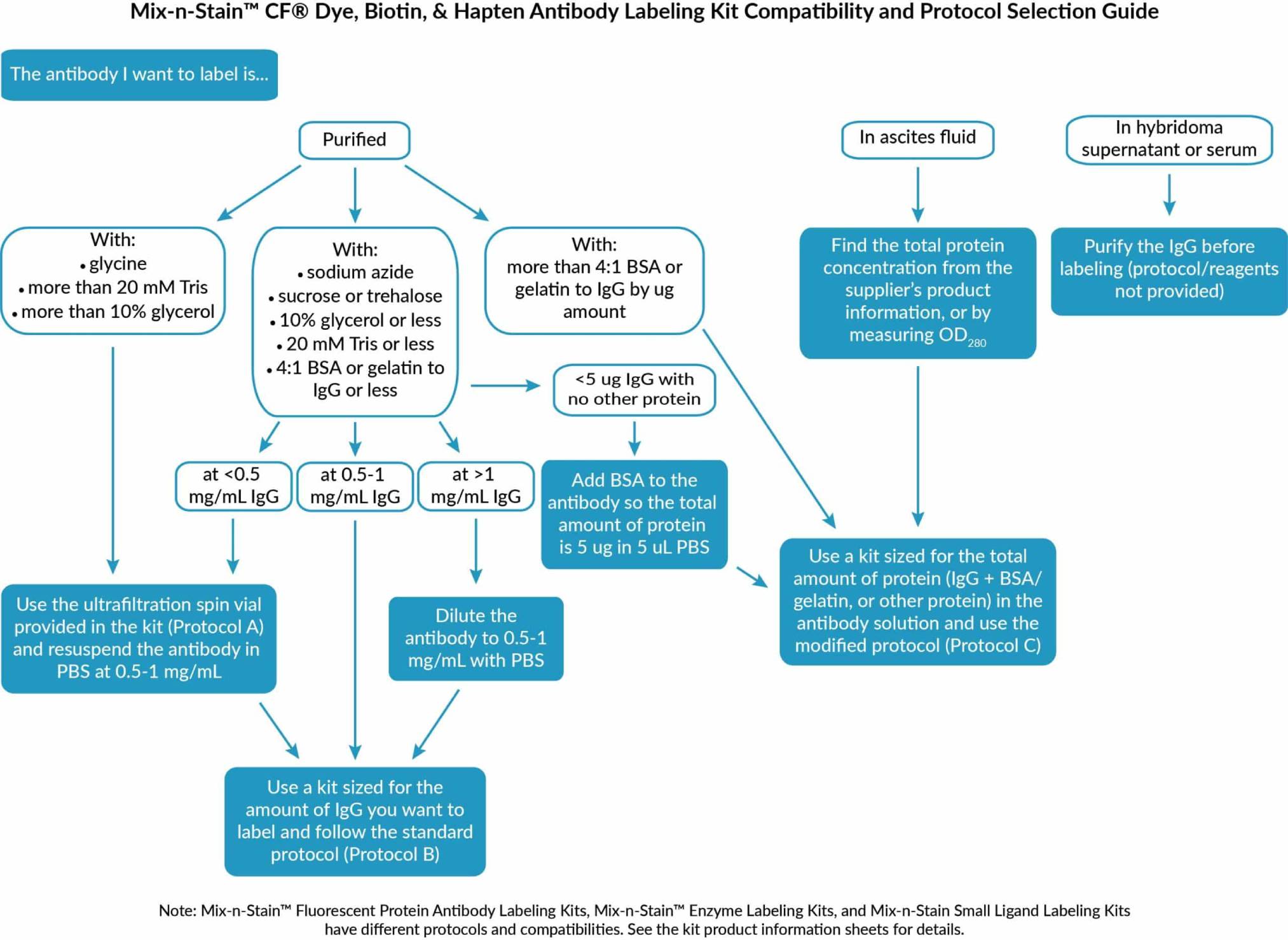
Mix-n-Stain™ CF®570 Antibody Labeling Kit, 1x(20-50ug) labeling

Cat# 92335
Size : 1kit
Brand : Biotium
Mix-n-Stain™ CF®570 Antibody Labeling Kit, 1x(20-50ug) labeling
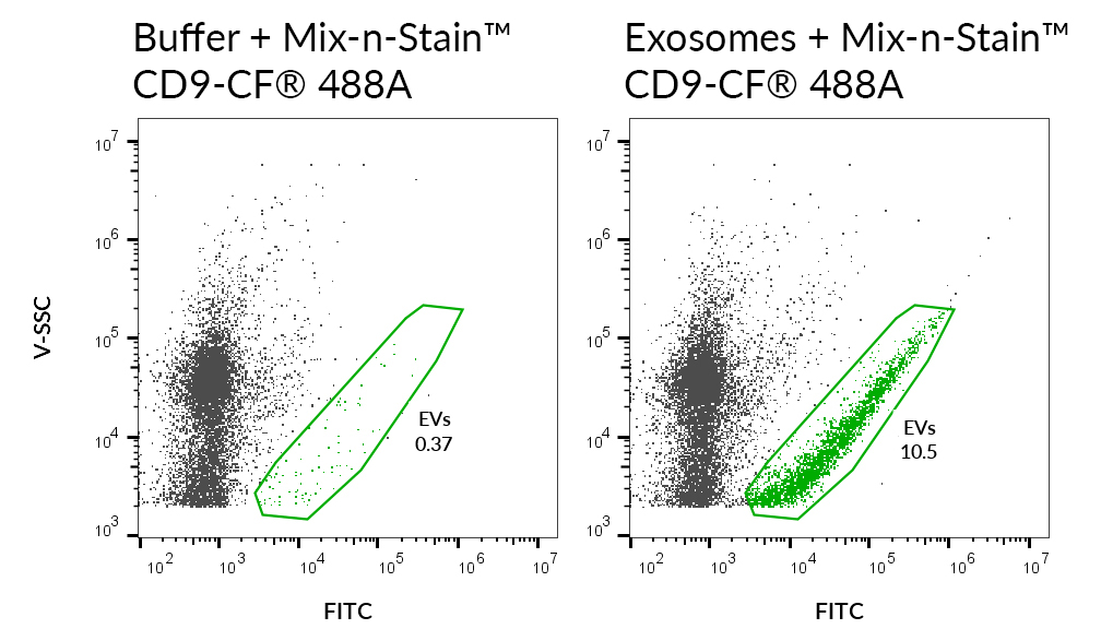 SEC-purified MCF-7-derived exosomes were stained with CD9 monoclonal antibody (CD9/2343) labeled with CF®488A using a Mix-n-Stain™ kit (right). Specific staining was seen, compared with the same antibody in buffer (left). Exosomes were detected on a CytoFLEX LX flow cytometer in the FITC channel.
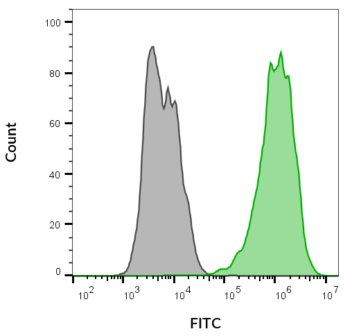 Flow cytometry of MCF-7 cells unstained (gray) or stained with CD9 monoclonal antibody (CD9/2343) labeled with CF®488A using a Mix-n-Stain™ kit (green).
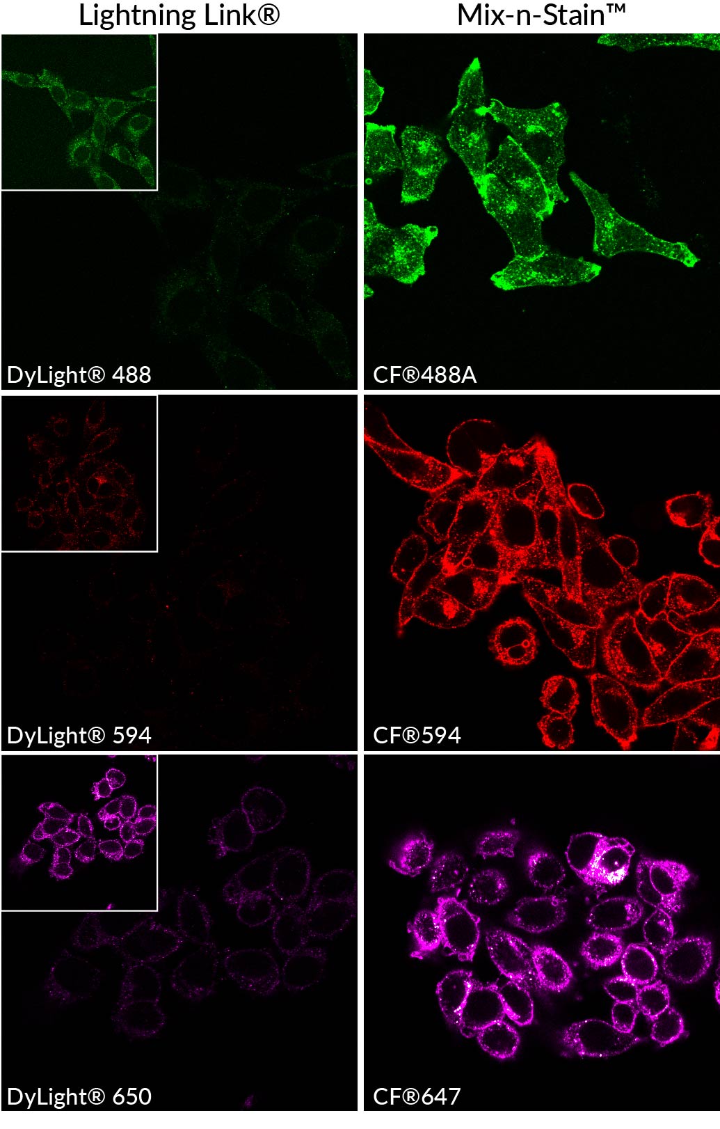 Mouse anti-transferrin receptor antibody from (endosome and plasma membrane marker) was labeled using Lightning-Link® Rapid DyLight® Conjugation Kits from Novus Biologicals (left) or Mix-n-Stain™ CF® Dye Antibody Labeling Kits (right) according to manufacturers’ instructions. The CF® dye conjugates show higher signal and more specific staining compared the DyLight®conjugates when imaged using the same settings, due to the superior brightness of CF® dyes. The insets show the same field of view imaged with a higher gain setting.
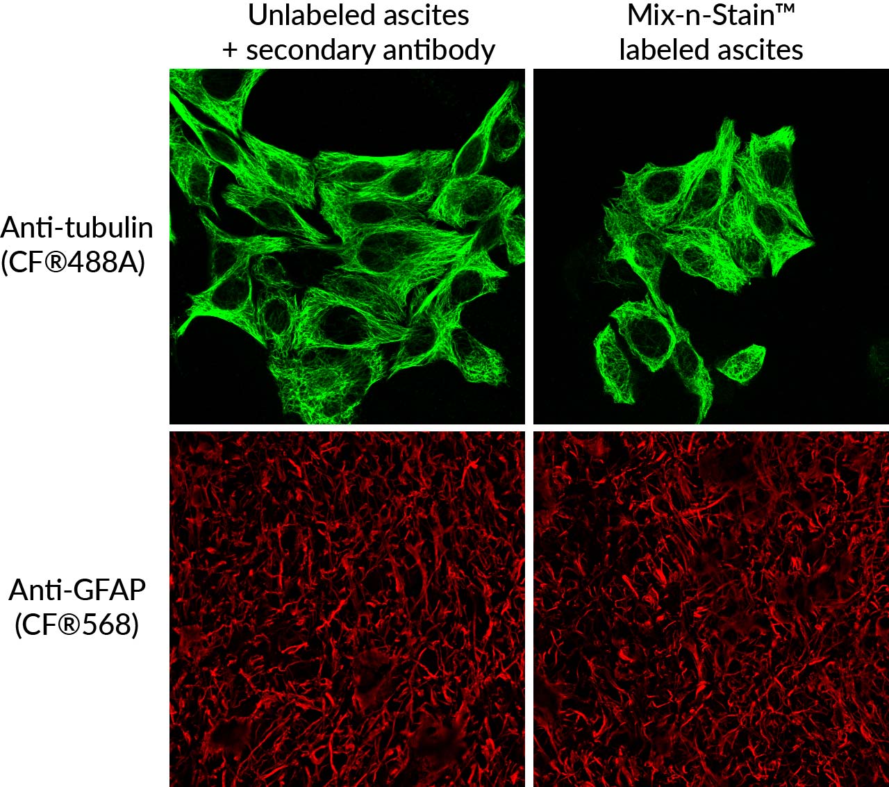 Mix-n-Stain™ labeling can be performed on unpurified antibodies in ascites fluid. Top: HeLa cells stained with anti-tubulin antibody in ascited, detected with CF®488A secondary antibody (left) or directly labeled using CF®488A Mix-n-Stain™ (right). Bottom: rat optic nerve cryosections stained with anti-GFAP (glial marker) detected with CF®568 secondary antibody or directly labeled with Mix-n-Stain™ CF®568. antibodies in Mix-n-Stain™ labeled ascites show comparable specificity to unlabeled antibodies detected using secondary antibody.
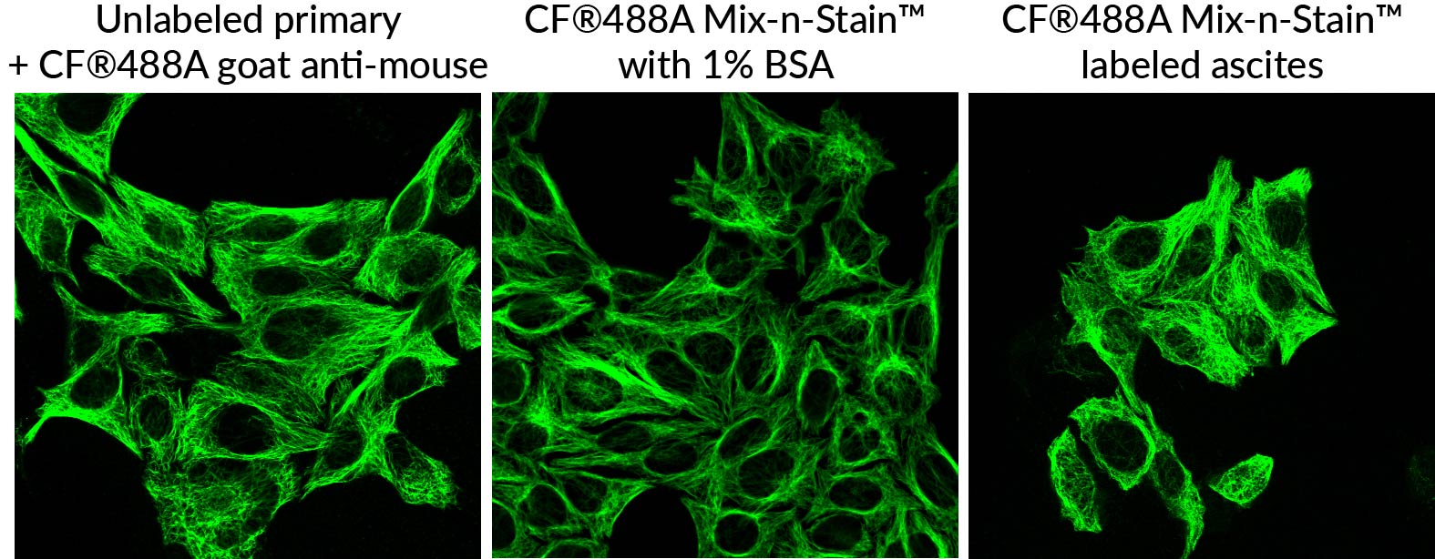 Monoclonal mouse anti-tubulin clone DM1A in ascites or purified in PBS with 1% BSA was labeled with CF®488A Mix-n-Stain using the modified protocol for total protein labeling. The Mix-n-Stain antibodies performed comparably in immunofluorescence staining of HeLa cells as unlabeled antibody (in ascites) detected with secondary antibody.
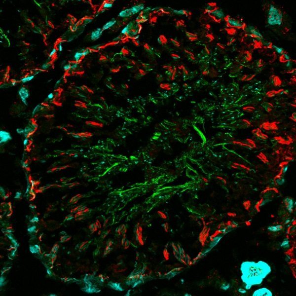 Rat testis with mouse anti-tubulin and CF®488A goat anti-mouse (min x rat) (green), CF®555 Mix-n-Stain labeled mouse anti-ZO1 (tight junctions, red) and CF®640R phalloidin (cyan).
  HeLa cells stained with CF640R Mix-n-Stain labeled mouse anti-tubulin antibody (red). Actin filaments are stained with CF488A phalloidin (green). Mounted in Everbrite Mounting Medium with DAPI (nuclei, blue).
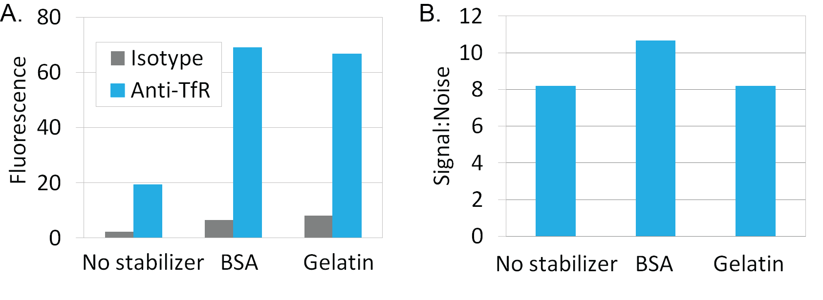 Mix-n-Stain™ labeling in the presence of 10-fold excess BSA or gelatin to IgG by weight using the modified Mix-n-Stain™ protocol. Mouse anti-transferrin receptor IgG or mouse IgG isotype control from BD Pharmingen were labeled using CF647 Mix-n- Stain™ kits. Intracellular staining of Jurkat cells was analyzed in channel FL3 of a BD FACSCalibur flow cytometer. Antibodies labeled in the presence of stabilizer showed comparable signal to noise ratio compared to conjugates labeled without stabilizer. A. Graph shows the geometric mean fluorescence of the cell populations. B. Graph shows signal to noise ratio (anti-TfR fluorescence/isotype fluorescence).
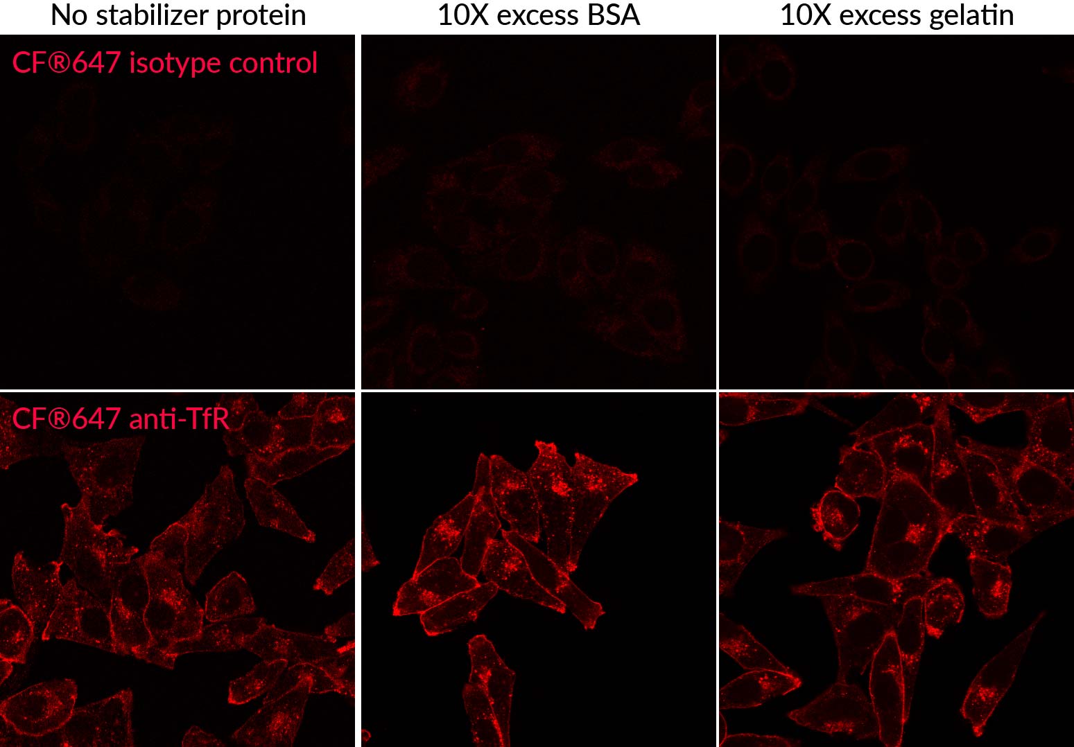 Comparison of Mix-n-Stain™ CF®647 labeling of anti-TfR antibody (endosome and cell surface marker) in HeLa cells without added protein compared to 1% BSA or 1% gelatin (10-fold excess) using the modified protocol for labeling in the presence of stabilizer protein. Labeling using the modified protocol gives comparable results as the standard protocol, with bright signal and minimal increase in non-specific background.
 SEC-purified MCF-7-derived exosomes were stained with CD9 monoclonal antibody (CD9/2343) labeled with CF®488A using a Mix-n-Stain™ kit (right). Specific staining was seen, compared with the same antibody in buffer (left). Exosomes were detected on a CytoFLEX LX flow cytometer in the FITC channel.Flow cytometry of MCF-7 cells unstained (gray) or stained with CD9 monoclonal antibody (CD9/2343) labeled with CF®488A using a Mix-n-Stain™ kit (green).Mouse anti-transferrin receptor antibody from (endosome and plasma membrane marker) was labeled using Lightning-Link® Rapid DyLight® Conjugation Kits from Novus Biologicals (left) or Mix-n-Stain™ CF® Dye Antibody Labeling Kits (right) according to manufacturers’ instructions. The CF® dye conjugates show higher signal and more specific staining compared the DyLight®conjugates when imaged using the same settings, due to the superior brightness of CF® dyes. The insets show the same field of view imaged with a higher gain setting.Mix-n-Stain™ labeling can be performed on unpurified antibodies in ascites fluid. Top: HeLa cells stained with anti-tubulin antibody in ascited, detected with CF®488A secondary antibody (left) or directly labeled using CF®488A Mix-n-Stain™ (right). Bottom: rat optic nerve cryosections stained with anti-GFAP (glial marker) detected with CF®568 secondary antibody or directly labeled with Mix-n-Stain™ CF®568. antibodies in Mix-n-Stain™ labeled ascites show comparable specificity to unlabeled antibodies detected using secondary antibody.Monoclonal mouse anti-tubulin clone DM1A in ascites or purified in PBS with 1% BSA was labeled with CF®488A Mix-n-Stain using the modified protocol for total protein labeling. The Mix-n-Stain antibodies performed comparably in immunofluorescence staining of HeLa cells as unlabeled antibody (in ascites) detected with secondary antibody.Rat testis with mouse anti-tubulin and CF®488A goat anti-mouse (min x rat) (green), CF®555 Mix-n-Stain labeled mouse anti-ZO1 (tight junctions, red) and CF®640R phalloidin (cyan). HeLa cells stained with CF640R Mix-n-Stain labeled mouse anti-tubulin antibody (red). Actin filaments are stained with CF488A phalloidin (green). Mounted in Everbrite Mounting Medium with DAPI (nuclei, blue).Mix-n-Stain™ labeling in the presence of 10-fold excess BSA or gelatin to IgG by weight using the modified Mix-n-Stain™ protocol. Mouse anti-transferrin receptor IgG or mouse IgG isotype control from BD Pharmingen were labeled using CF647 Mix-n- Stain™ kits. Intracellular staining of Jurkat cells was analyzed in channel FL3 of a BD FACSCalibur flow cytometer. Antibodies labeled in the presence of stabilizer showed comparable signal to noise ratio compared to conjugates labeled without stabilizer. A. Graph shows the geometric mean fluorescence of the cell populations. B. Graph shows signal to noise ratio (anti-TfR fluorescence/isotype fluorescence).Comparison of Mix-n-Stain™ CF®647 labeling of anti-TfR antibody (endosome and cell surface marker) in HeLa cells without added protein compared to 1% BSA or 1% gelatin (10-fold excess) using the modified protocol for labeling in the presence of stabilizer protein. Labeling using the modified protocol gives comparable results as the standard protocol, with bright signal and minimal increase in non-specific background.