EpCAM (epithelial cell adhesion molecule; CD326; EPCAM/TACSTD1) is a 35–40 kDa type I transmembrane glycoprotein expressed predominantly on the basolateral membrane of simple, pseudostratified, and transitional epithelial cells. Physiologically, EpCAM is highly expressed in the intestinal epithelium and is also present in pancreatic, biliary, and colorectal mucosal epithelium. Beyond its classical role in calcium-independent cell–cell adhesion, EpCAM contributes to epithelial homeostasis, proliferation, differentiation, stem cell maintenance, and intracellular signaling through interactions with the WNT/β-catenin pathway and downstream targets including c-Myc. Aberrant overexpression in epithelial malignancies has established EpCAM as an important biomarker in gastrointestinal cancer diagnostics and translational oncology research.
Biological Significance of EpCAM
- Mediates calcium-independent epithelial cell adhesion and contributes to the maintenance of tissue architecture.
- Regulates epithelial proliferation, differentiation, and tissue regeneration.
- Participates in intracellular signaling following regulated intramembrane proteolysis and release of the EpICD intracellular domain.
- Is associated with epithelial stem cell phenotypes and tumor progression in gastrointestinal malignancies.
- Shows particularly high physiological expression in intestinal epithelium and is also expressed in pancreatobiliary epithelial cells.
Diagnostic Utility of EpCAM in Gastrointestinal Pathology
- Serves as a sensitive marker of epithelial differentiation in gastrointestinal adenocarcinomas.
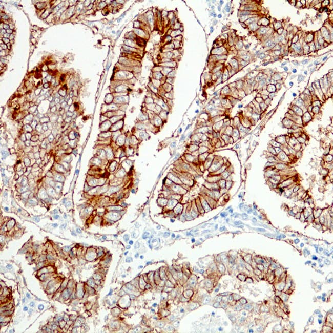
- Strong membranous staining is commonly observed in colorectal adenocarcinoma, gastric adenocarcinoma, pancreatic ductal adenocarcinoma, cholangiocarcinoma, and many metastatic gastrointestinal carcinomas.
- Is useful in distinguishing epithelial carcinomas from many mesenchymal and lymphoid neoplasms and may contribute to the differential diagnosis with selected neuroendocrine neoplasms when interpreted alongside lineage-specific markers.
- Assists in identifying metastatic adenocarcinoma deposits in lymph nodes, liver, and serosal surfaces.
- Complements markers such as CK7, CK20, CDX2, SATB2, MUC1, and MUC2 for tumour classification and determination of the primary site. EpCAM expression should be interpreted cautiously in poorly differentiated carcinomas and tumours undergoing epithelial–mesenchymal transition, where expression may be reduced or lost.
Key Features of CE-IVD Anti-EpCAM Antibodies for IHC
- CE-marked for in vitro diagnostic use on formalin-fixed paraffin-embedded (FFPE) tissues.
- Optimized for both automated and manual immunohistochemistry workflows.
- Typically demonstrate crisp membranous staining with minimal background signal.
- Suitable for gastrointestinal tumour typing and metastatic tumour characterization.
- Available in ready-to-use and concentrated formats compatible with major staining platforms.