


DNMT3A antibody


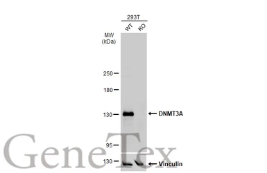
Wild-type (WT) and DNMT3A knockout (KO) 293T cell extracts (30 μg) were separated by 5% SDS-PAGE, and the membrane was blotted with DNMT3A antibody (GTX129125) diluted at 1:1000. The HRP-conjugated anti-rabbit IgG antibody (GTX213110-01) was used to detect the primary antibody.
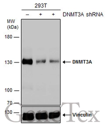
Non-transfected (–) and transfected (+) 293T whole cell extracts (30 μg) were separated by 5% SDS-PAGE, and the membrane was blotted with DNMT3A antibody (GTX129125) diluted at 1:500. The HRP-conjugated anti-rabbit IgG antibody (GTX213110-01) was used to detect the primary antibody.
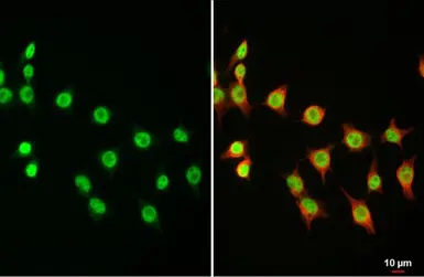
DNMT3A antibody detects DNMT3A protein at nucleus by immunofluorescent analysis.
Sample: 293T cells were fixed in 4% paraformaldehyde at RT for 15 min.
Green: DNMT3A stained by DNMT3A antibody (GTX129125) diluted at 1:1000.
Red: alpha Tubulin, a cytoskeleton marker, stained by alpha Tubulin antibody [GT114] (GTX628802) diluted at 1:1000.
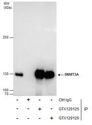
Immunoprecipitation of DNMT3A protein from 293T whole cell extracts using 5 μg of DNMT3A antibody (GTX129125) or DNMT3A antibody (GTX129126).
Western blot analysis was performed using DNMT3A antibody (GTX129125) diluted at 1:2000.
EasyBlot anti-Rabbit IgG (GTX221666-01) was used as a secondary reagent.
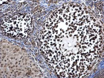
DNMT3A antibody detects DNMT3A protein at nucleus on mouse ovary by immunohistochemical analysis.
Sample: Paraffin-embedded mouse ovary.
DNMT3A antibody (GTX129125) dilution: 1:500.
Antigen Retrieval: Trilogy™ (EDTA based, pH 8.0) buffer, 15min
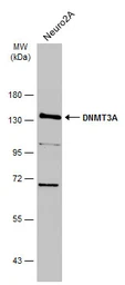
Whole cell extract (30 μg) was separated by 7.5% SDS-PAGE, and the membrane was blotted with DNMT3A antibody (GTX129125) diluted at 1:1000. The HRP-conjugated anti-rabbit IgG antibody (GTX213110-01) was used to detect the primary antibody.
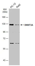
Various whole cell extracts (30 μg) were separated by 7.5% SDS-PAGE, and the membrane was blotted with DNMT3A antibody (GTX129125) diluted at 1:1000. The HRP-conjugated anti-rabbit IgG antibody (GTX213110-01) was used to detect the primary antibody.
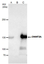
DNMT3A antibody immunoprecipitates DNMT3A protein in IP experiments.
IP samples: 293T nuclear extract
A. 50 μg 293T nuclear extract
B. Control with 4 μg of preimmune Rabbit IgG
C. Immunoprecipitation of DNMT3A protein by 4 μg DNMT3A antibody (GTX129125)
5 % SDS-PAGE
The immunoprecipitated DNMT3A protein was detected by DNMT3A antibody (GTX129125) diluted at 1:2000.
[EasyBlot anti-rabbit IgG (GTX221666-01) was used as a secondary reagent]
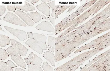
DNMT3A antibody detects DNMT3A protein at nucleus by immunohistochemical analysis.Sample: Paraffin-embedded mouse tissue.DNMT3A stained by DNMT3A antibody (GTX129125) diluted at 1:500.Antigen Retrieval: Citrate buffer, pH 6.0, 15 min
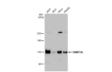
Various whole cell extracts (30 μg) were separated by 5% SDS-PAGE, and the membrane was blotted with DNMT3A antibody (GTX129125) diluted at 1:2000. The HRP-conjugated anti-rabbit IgG antibody (GTX213110-01) was used to detect the primary antibody.
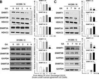
The data was published in the journal Oncotarget in 2018. PMID: 29416619
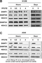
The data was published in the journal Oncotarget in 2018. PMID: 29416619
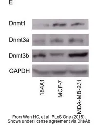
The data was published in the journal PLoS One in 2015. PMID: 25928539