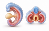

Embryonic Folding & Body Plan Formation
Embryonic folding is a fundamental morphogenetic process that transforms the relatively flat trilaminar embryonic disc into a three-dimensional, approximately cylindrical embryo. In humans, major body folding occurs during Carnegie stages 9–13, in parallel with neurulation, somitogenesis, and the onset of organogenesis. Folding reorganizes the three germ layers and establishes the spatial relationships required for subsequent organ development.
Mechanisms of Embryonic Folding
Two coordinated components contribute to this transformation:
-
Cephalocaudal folding bends the embryo along its longitudinal axis, producing cranial and caudal folds and incorporating portions of the endoderm-lined yolk sac into the developing foregut and hindgut.
-
Lateral folding brings the lateral embryonic margins ventrally toward the midline, contributing to ventral body-wall closure and incorporation of yolk-sac endoderm into the primitive gut. The midgut remains temporarily connected to the yolk sac through the vitelline duct.
Embryonic folding reflects the interaction of differential tissue growth, tissue geometry, mechanical constraints, and active cellular behaviors. Rapid growth of the neural tissues and somites, together with changes in the surrounding mesoderm and extraembryonic tissues, contributes to the deformation of the embryonic disc.
Tissue Mechanics and Remodeling
Folding is influenced by tissue-level mechanical properties, including differential growth, tissue stiffness, cell–cell and cell–extracellular-matrix adhesion, and cytoskeletal contractility. These properties interact with regional tissue geometry and developmental patterning to generate the deformations associated with body folding. ECM composition and remodeling can influence tissue mechanics and morphogenesis, although their contributions vary between tissues and developmental processes.
Germ-Layer Reorganization and Tubulogenesis
Folding internalizes part of the endoderm-lined yolk sac to form the primitive gut tube, while the lateral plate mesoderm separates into somatic and splanchnic components around the intraembryonic coelom. The coelomic space is subsequently partitioned and remodeled to form the definitive pericardial, pleural, and peritoneal cavities.
The resulting three-dimensional arrangement places mesoderm between ectoderm and endoderm and establishes the anatomical relationships from which the nervous system, body wall, cardiovascular system, gut, and other organ systems subsequently develop.
Molecular and Cellular Regulation
Embryonic folding occurs within a broader developmental program involving molecular patterning and tissue mechanics. Signaling pathways including WNT/planar cell polarity, FGF, BMP, NODAL, SHH, and retinoic acid participate in the patterning and morphogenesis of the early embryo, although their roles are not equivalent or necessarily direct drivers of body folding. Cellular mechanisms relevant to morphogenesis during this period include differential growth, cell-shape changes, polarized cell intercalation, actomyosin contractility, and remodeling of cell–cell and cell–ECM interactions.
Body-Plan Formation
Folding transforms the previously established embryonic axes into a three-dimensional body architecture and reorganizes tissues according to their cranial–caudal, dorsal–ventral, and left–right positional relationships. It positions the developing nervous system, somites, gut, cardiovascular structures, and body wall relative to one another, providing the anatomical framework for subsequent organogenesis.
Pre-existing left–right patterning is subsequently manifested through asymmetric morphogenesis, including directional cardiac looping and the development of visceral asymmetries.