CA9 Polyclonal Antibody

Referencia E-AB-64788-200
embalaje : 200uL
Marca : Elabscience
 Resources
-
-
-
- +1
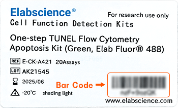
For research use only.
| Verified Samples | Verified Samples in WB: Mouse small intestine, Rat small intestine Verified Samples in IHC: Human stomach, Human gastric cancer Verified Samples in IF: Hela |
| Dilution | WB 1:500-1:2000, IHC 1:50-1:200, IF 1:50-1:200 |
| Isotype | IgG |
| Host | Rabbit |
| Reactivity | Human, Mouse, Rat |
| Applications | WB, IHC, IF |
| Clonality | Polyclonal |
| Immunogen | Recombinant fusion protein of human CA9 (NP_001207.2). |
| Abbre | CA9 |
| Synonyms | CA9, CAIX, MN |
| Swissprot | |
| Calculated MW | 49 kDa |
| Observed MW | 58 kDa The actual band is not consistent with the expectation.
Western blotting is a method for detecting a certain protein in a complex sample based on the specific binding of antigen and antibody. Different proteins can be divided into bands based on different mobility rates. The mobility is affected by many factors, which may cause the observed band size to be inconsistent with the expected size. The common factors include: 1. Post-translational modifications: For example, modifications such as glycosylation, phosphorylation, methylation, and acetylation will increase the molecular weight of the protein. 2. Splicing variants: Different expression patterns of various mRNA splicing bodies may produce proteins of different sizes. 3. Post-translational cleavage: Many proteins are first synthesized into precursor proteins and then cleaved to form active forms, such as COL1A1. 4. Relative charge: the composition of amino acids (the proportion of charged amino acids and uncharged amino acids). 5. Formation of multimers: For example, in protein dimer, strong interactions between proteins can cause the bands to be larger. However, the use of reducing conditions can usually avoid the formation of multimers. If a protein in a sample has different modified forms at the same time, multiple bands may be detected on the membrane. |
| Cellular Localization | Nucleus. Nucleus, nucleolus. Cell membrane. Cell projection, microvillus membrane. Found on the surface microvilli and in the nucleus, particularly in nucleolus. |
| Concentration | 1 mg/mL |
| Buffer | Phosphate buffered solution, pH 7.4, containing 0.05% stabilizer and 50% glycerol. |
| Purification Method | Affinity purification |
| Research Areas | Cancer, Cardiovascular, Metabolism, Signal Transduction |
| Conjugation | Unconjugated |
| Storage | Store at -20°C Valid for 12 months. Avoid freeze / thaw cycles. |
| Shipping | The product is shipped with ice pack,upon receipt,store it immediately at the temperature recommended. |
| background | Carbonic anhydrases (CAs) are a large family of zinc metalloenzymes that catalyze the reversible hydration of carbon dioxide. They participate in a variety of biological processes, including respiration, calcification, acid-base balance, bone resorption, and the formation of aqueous humor, cerebrospinal fluid, saliva, and gastric acid. They show extensive diversity in tissue distribution and in their subcellular localization. CA IX is a transmembrane protein and is one of only two tumor-associated carbonic anhydrase isoenzymes known. It is expressed in all clear-cell renal cell carcinoma, but is not detected in normal kidney or most other normal tissues. It may be involved in cell proliferation and transformation. This gene was mapped to 17q21.2 by fluorescence in situ hybridization, however, radiation hybrid mapping localized it to 9p13-p12. |
-
IF:{{item.impact}}
Journal:{{item.journal}} ({{item.year}})
DOI:{{item.doi}}Reactivity:{{item.species}}
Sample Type:{{item.sample_type}}
-
Q{{(FAQpage.currentPage - 1)*pageSize+index+1}}:{{item.name}}
No files available. Please enter the correct identification code in the search area to query the relevant documents.

| Title | Status | Valid until | Action | |
|---|---|---|---|---|
| {{v.文件名}} | {{v.status_str}} | {{v.timestamp_str}} | Download | / |
*Note:Please download the relevant files within the validity period. After expiration, file preview and download will not be available on the page.
Videos
Resources
Contact with Us
Houston, Texas, 77079, USA
