

CellBrite® Fix 488 Membrane Labeling Kit, Trial Size

Referencia 30090-T
embalaje : 1kit
Marca : Biotium
CellBrite® Fix Membrane Stains
Unique fluorogenic membrane dyes that covalently stain the plasma membrane in live cells, and can withstand both fixation and detergent permeabilization.
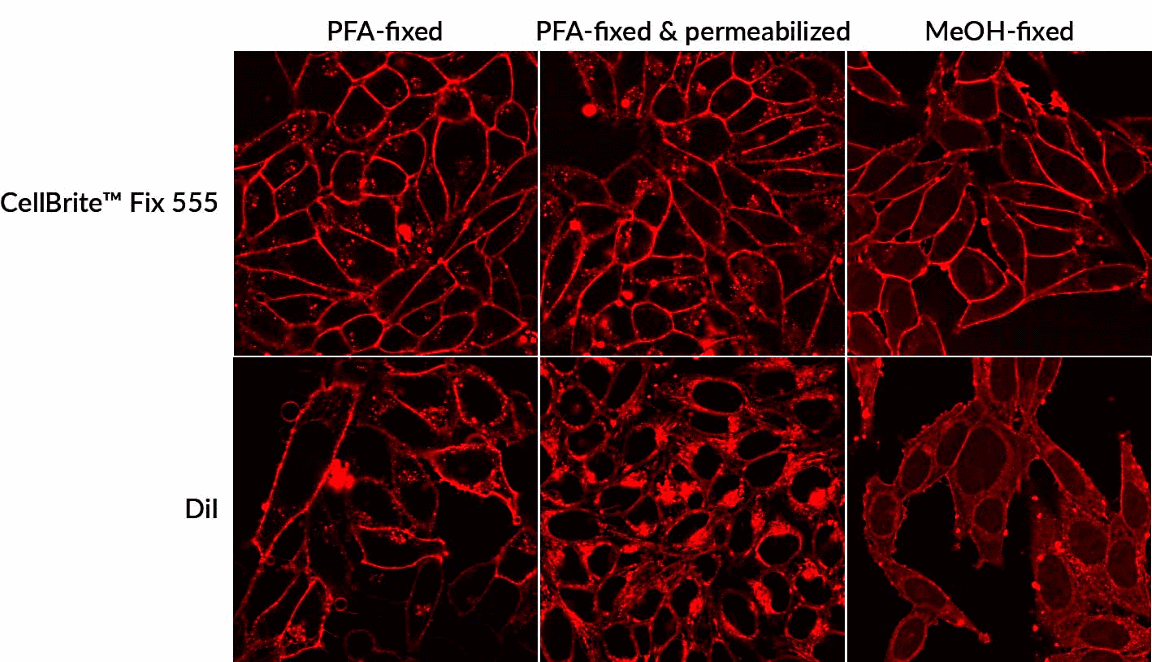 Comparison of traditional carbocyanine dye and CellBrite™ Fix before and after fixation in HeLa cells. CellBrite™ Fix 555 gave more even surface staining and was better retained after fixation/permeabilization compared to DiI.
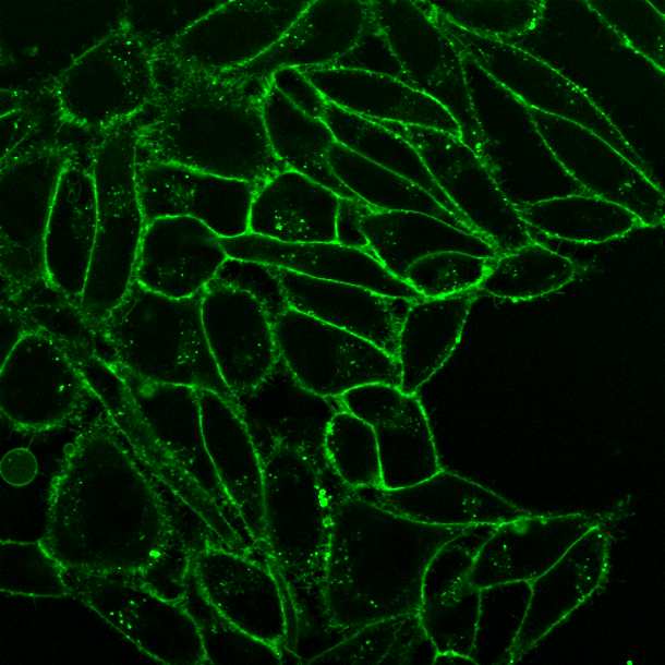 Live HeLa cells stained with 1X CellBrite™ Fix 488 for 15 minutes at 37°C.
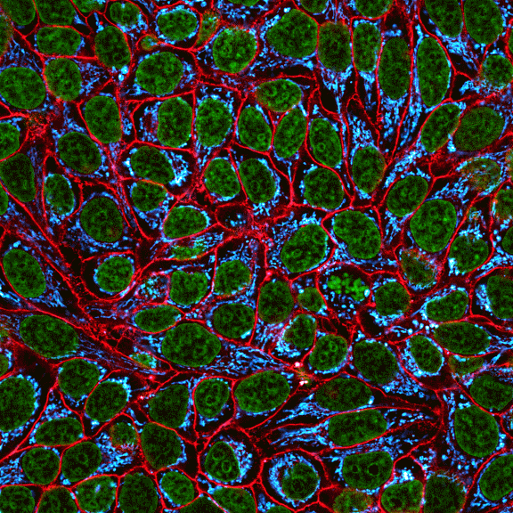 HeLa cell membranes were stained with CellBrite Fix 555 (red) for 15 minutes at 37°C, then fixed with MeOH. Mitochondria were stained with CF®640R Anti-Mitochondrial Marker clone 111-3 (cyan) and nuclei were stained with NucSpot® 470 (green).
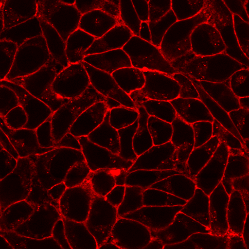 HeLa cells labeled with CellBrite™ Fix for 30 minutes at room temperature, then fixed with paraformaldehyde and permeabilized with Triton™ X-100.
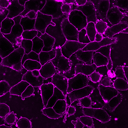 HeLa cells labeled with CellBrite Fix 640 for 30 minutes at room temperature, then fixed with methanol.
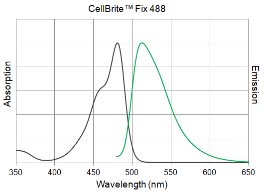 Absorption and emission of CellBrite™ Fix 488 dye in water.
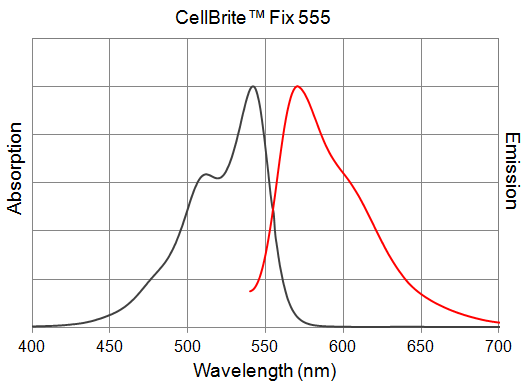 Absorption and emission of CellBrite™ Fix 555 dye in water.
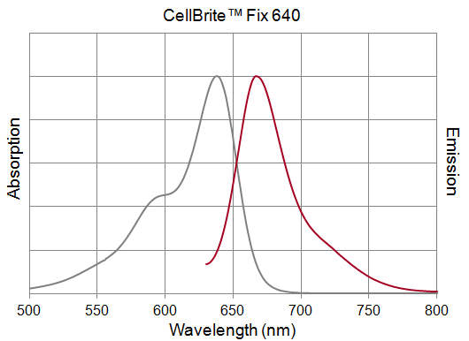 Absorption and emission of CellBrite™ Fix 640 dye in water.
Product Description
CellBrite® Fix dyes are unique fluorogenic membrane dyes that covalently stain the plasma membrane in live cells, and can withstand both fixation and detergent permeabilization.
- The only membrane dyes that withstand fixation and permeabilization
- Bright, uniform cell surface staining in 15 minutes
- Available with green, red and far-red fluorescence
- CellBrite® Fix 488 validated for super-resolution imaging of exosomes by SIM (Ref. 1)
- Each vial makes 20 uL of 1000X dye
Unique Fixable Membrane Stains
CellBrite® Fix dyes are a series of proprietary fluorophores developed by Biotium to rapidly stain the outer plasma membranes of live cells. They are unique among membrane stains in that their surface staining can withstand permeabilization and methanol fixation, allowing plasma membrane staining to be combined with intracellular staining with antibodies. Other membrane dyes like DiO, DiI, Vybrant® membrane dyes, CellMask™, CellVue®, or PKH dyes can be fixed with formaldehyde. But they are not compatible with detergent permeabilization or methanol fixation, because these treatments extract lipophilic dyes from membranes. Unlike lectins such as WGA, which bind specific targets that may vary between cell types, CellBrite® Fix dyes are general membrane stains.
CellBrite® Fix dyes are designed to accumulate at the cell membrane, where they become covalently attached to membrane proteins. As a result, surface staining is well-retained after permeabilization or methanol fixation, with only a slight increase in intracellular fluorescence compared to formaldehyde fixation alone. CellBrite® Fix dyes are only weakly fluorescent in aqueous media but become intensely fluorescent upon membrane staining. This fluorogenic property of the dyes makes the staining very specific with low background. Due to their better water solubility, CellBrite® Fix dyes yield much more uniform staining compared to lipophilic carbocyanine dyes like DiO and DiI. The dyes are non-cytotoxic and do not readily transfer between cells.
CellBrite® Fix dyes also can be used to stain yeast and bacteria (gram-positive or gram-negative). See our Cellular Stains Table for more information on how our dyes stain various organisms.
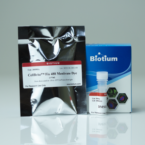
CellBrite® Fix Membrane Stains are provided in lyophilized format. A vial of anhydrous DMSO is included for dye reconstitution. After reconstitution, each vial yields 20 uL of 1000X dye (download the product protocol for more information). The dyes are available with green, red, and far-red fluorescence. We also offer MemBrite® Fix Cell Surface Staining Kits. with a wide selection of dye colors, including super-resolution compatible options. For more information, see Frequently Asked Questions.
Tips for Success
Note that CellBrite® Fix dyes stain dead cell more intensely than live cells. Please see our Tech Tip: Five Steps for Success with Membrane and Surface Stains for tips on staining and imaging (step 5) with CellBrite® Fix.
Find the Right Stain for Your Application
CellBrite® Fix dyes must be used to stain live cells before fixation. They cannot be used to stain cells that are already fixed (the dyes primarily label intracellular membranes in fixed cells). Our original CellBrite® Cytoplasmic Membrane Dyes can be used to stain cells after fixation and permeabilization, see our Tech Tip: Combining Lipophilic Membrane Dyes with Immunofluorescence. To find the right stain for your application, see our Membrane & Cell Surface Stains Comparison, or download our Membrane & Surface Stains Brochure.
CellBrite® Fix dyes are designed to be fixed shortly after staining. With prolonged dye incubation, or if cells are cultured after staining, the dyes will be internalized by endocytosis, resulting in labeling of intracellular vesicles. By 24 hours after staining, most of the dye will be localized inside the cell, not on the cell surface. For long-term visualization of cell boundaries in culture, we recommend our CellBrite® Steady Membrane Staining Kits. These kits include unique fluorescent membrane probes that retain cell surface staining in live cell cultures for 24 hours or longer. The kits also include an optional CellBrite® Steady Enhancer solution which masks intracellular signal for even greater specificity of cell boundaries.
CellBrite® Fix 488 has been reported to stain exosomes for super-resolution imaging by SIM (Ref. 1; see the Highlighted Citation). However, we find that CellBrite® Fix dyes do not efficiently stain isolated exosomes, instead we recommend using our ExoBrite™ EV Membrane Staining Kits for membrane staining of exosomes. See all of our recommended stains for Exosome & EV Labeling.
Note: CellBrite® Fix dyes carry an overall net positive charge. We have had customers report that they may be permeant to mechanotransduction or other cation channels on nerve cells, similar to cationic styryl dyes such as FM®1-43 (see J Neurosci (2013) 23(10), 4054), leading to significant intracellular accumulation or staining. Our MemBrite® Fix dyes are not positively charged, and therefore should not have this issue.
See also our GlycoLiner™ Cell Surface Glycoprotein Labeling Kits designed for covalent labeling of glycoproteins on the cell surface of live cells. GlycoLiner™ also has significantly less cytoplasmic background in dead cells than CellBrite® Fix or MemBrite® Fix stains, providing easier imaging of cell surfaces.
Watch our video where Technical Applications Scientist II, Jacqueline Steenhuis PhD answers your top questions about Biotium's various membrane stains for fluorescence microscopy.
For additional support or product recommendations, contact us at [email protected].
CellBrite® Fix Ordering Information
| Dye | Abs/Em | Size | Catalog number |
|---|---|---|---|
| CellBrite® Fix 488 | 480/513 nm | Trial size (1 vial) | 30090-T |
| Set of 5 vials | 30090 | ||
| CellBrite® Fix 555 | 542/571 nm | Trial size (1 vial) | 30088-T |
| Set of 5 vials | 30088 | ||
| CellBrite® Fix 640 | 638/667 nm | Trial size (1 vial) | 30089-T |
| Set of 5 vials | 30089 |
Technology

Tech Tips

Product Attributes
Size
Probe cellular localization
For live or fixed cells
Assay type/options
Fixation options
Colors
Documents, Protocols, SDS and COA

Protocols
COA
Request the Certificate of AnalysisReferences
Download a list of curated CellBrite® and MemBrite® references.
Citations
Download a list of curated CellBrite® and MemBrite® references.
 Powered by Bioz