

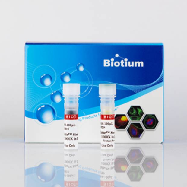
CellBrite® Steady 488 Membrane Staining Kit, 100 Labelings

Referencia 30106-T
embalaje : 1kit
Marca : Biotium
CellBrite® Steady Membrane Staining Kits
Cell surface staining kits with fluorescent membrane dye plus enhancer for imaging live cell surface for several hours to days.
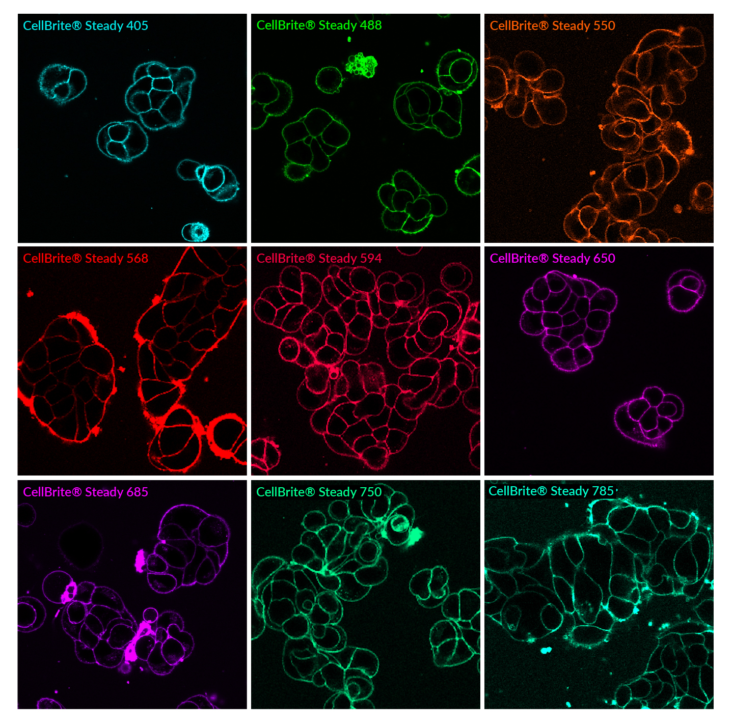 Live MCF7 cells stained with CellBrite® Steady dyes for 30 minutes and imaged by confocal microscopy without a wash step.
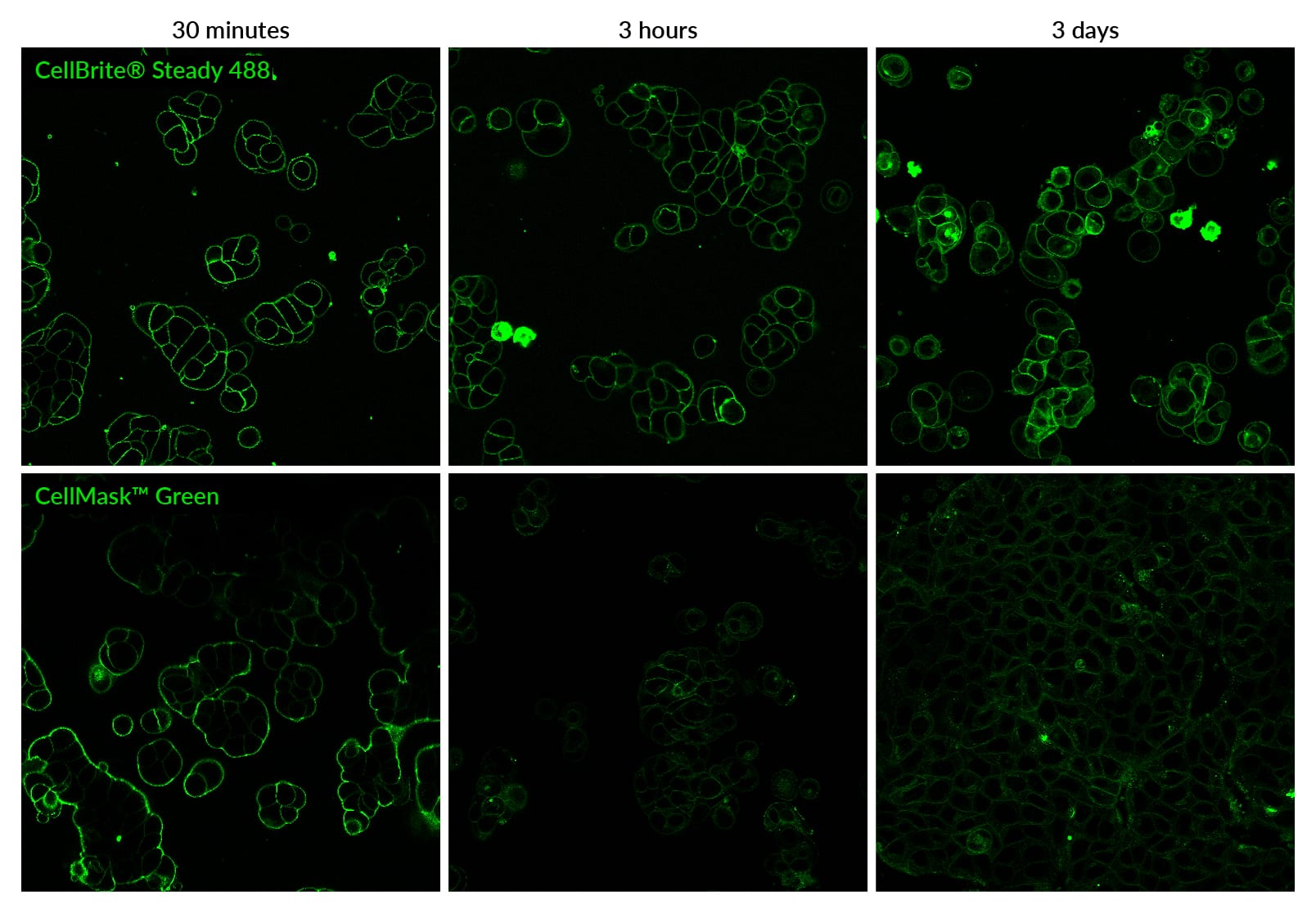 CellBrite® Steady staining is more stable than CellMask™ Green for real-time live cell imaging. Live MCF7 cells were stained with 1X CellBrite™ Steady 488 with CellBrite® Steady Enhancer in complete culture medium at 37°C without a wash step. Cells were cultured for 72 hours after staining and imaged at the time points shown. For CellMask™ Green staining, cell were stained according to the manufacturer's recommendations. Cells were stained with dye diluted 1:1000 in HBSS buffer for 10 minutes at 37°C, then rinsed in buffer, and then fresh culture medium was added. Cells were cultured for 72 hours and imaged at the time points shown.
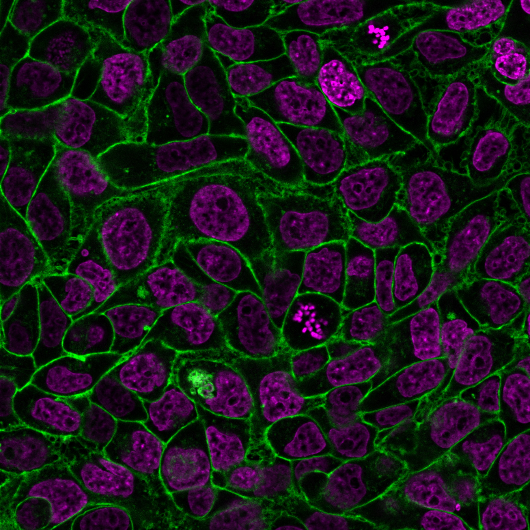 Live HeLa cells co-stained with CellBrite™ Steady 488 (green) with Enhancer and NucSpot® Live 650 (magenta) for 45 minutes. Cells were imaged by confocal microscopy without a wash step.
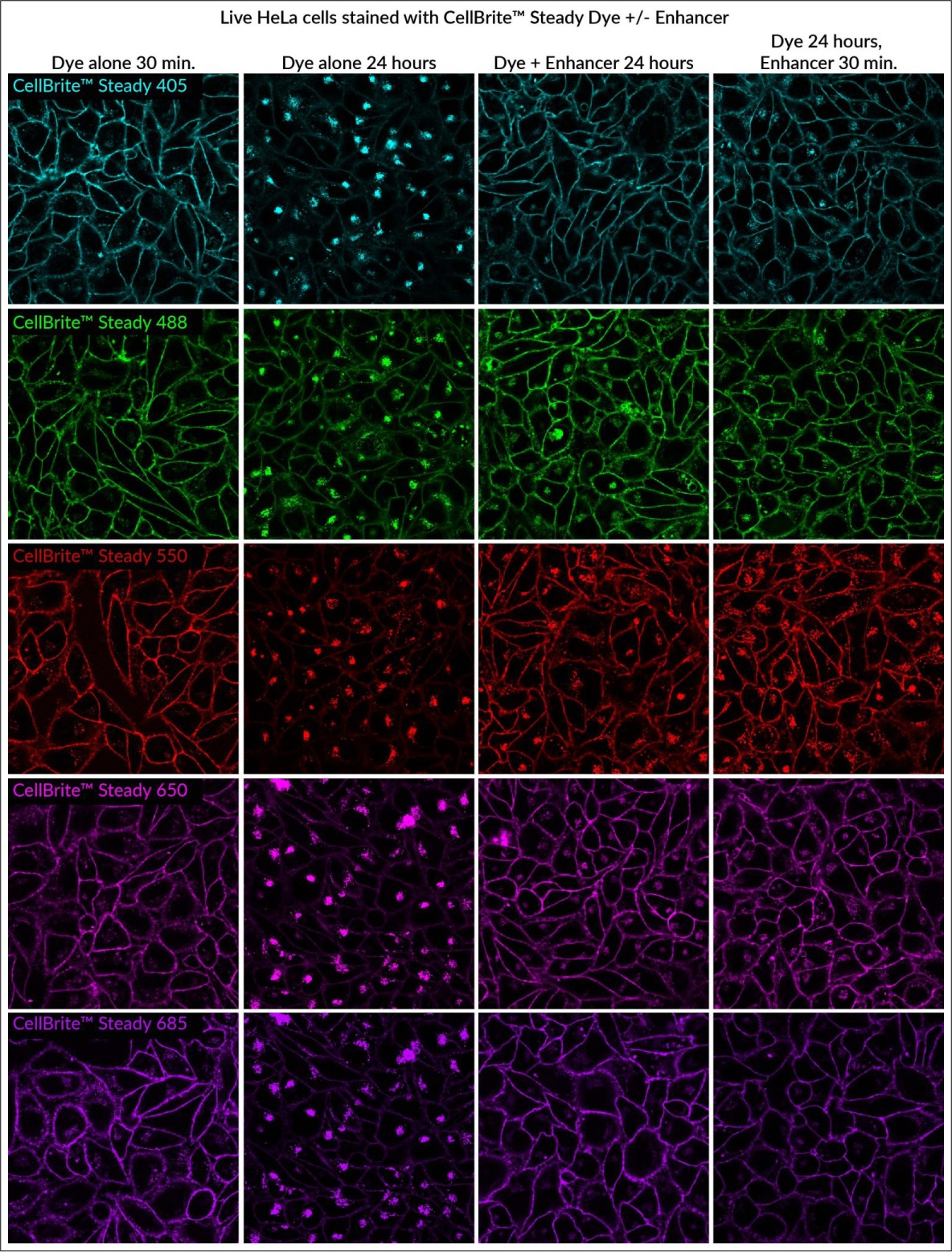 Comparison of live cell staining with CellBrite™ Steady Dyes with or without Enhancer. Live HeLa cells were stained for the indicated times. Over 24 hours, the dyes equilibrate between the plasma membrane and intracellular compartments. Co-incubating or post-treating with Enhancer masks intracellular staining, allowing selective imaging of cell boundaries. Cells were imaged by confocal microscopy without a wash step.
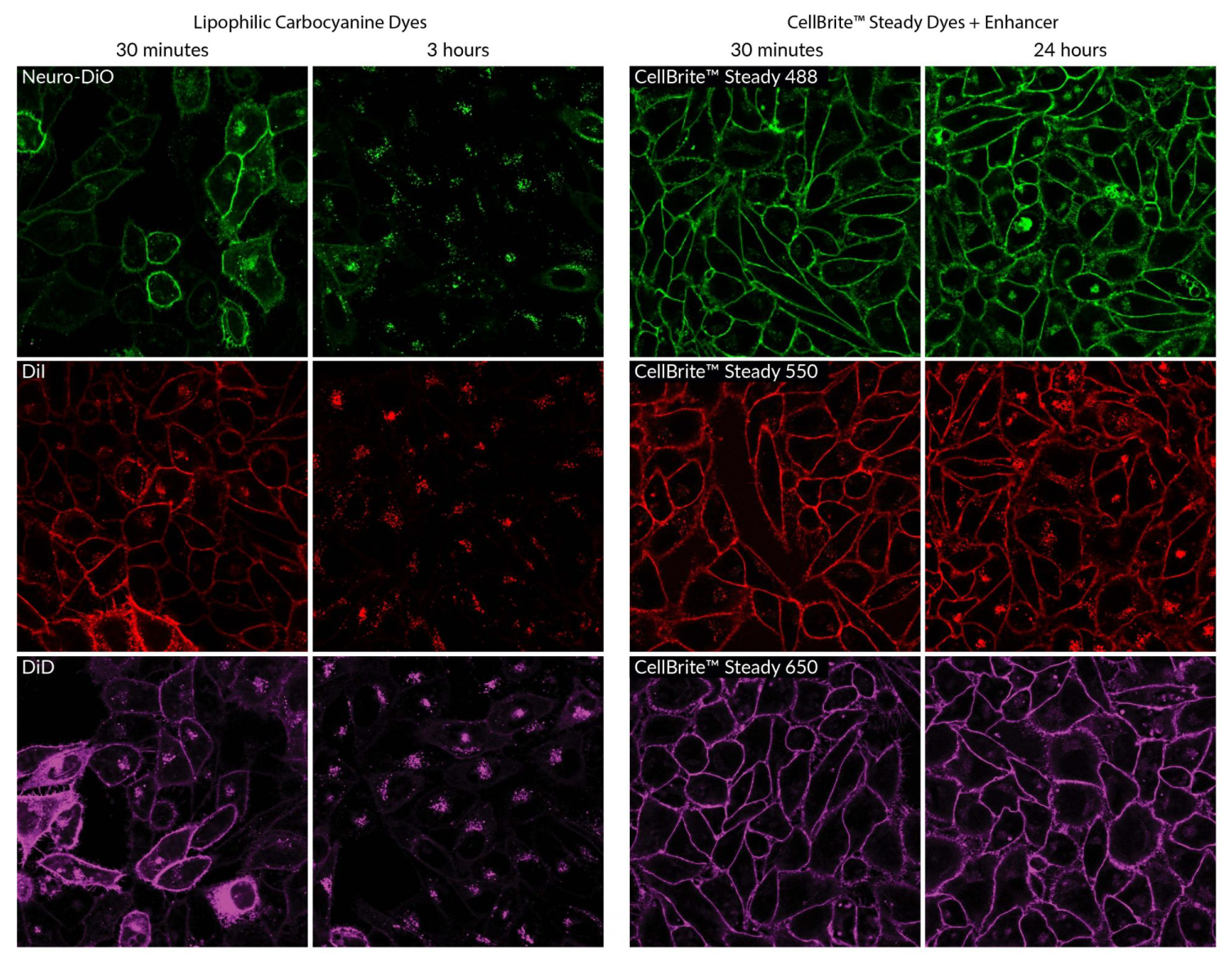 Live HeLa cells stained with the lipophilic carbocyanine dyes Neuro-DiO, DiI, or DiD or CellBrite® Steady Membrane Dyes and Enhancer. Lipophilic carbocyanine dyes are rapidly internalized into intracellular compartments, so surface staining must be imaged within short time frames. CellBrite® Steady Dyes stain cell membranes evenly, and surface staining can be clearly imaged for 24 hours with the use of Enhancer.
 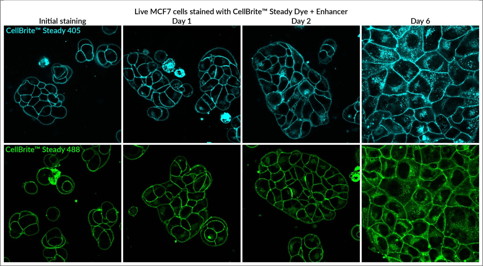 Long-term staining with CellBrite™ Steady Dyes and Enhancer. MCF7 cells stained with 1X CellBrite™ Steady 405 or CellBrite™ Steady 488 dye, co-incubated with 0.2X Enhancer for up to six days. Cells were imaged by confocal microscopy without a wash step.
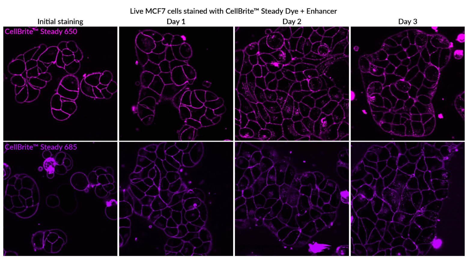 Live MCF7 cells stained with 1X CellBrite™ Steady 650 or CellBrite™ Steady 685 dye, co-incubated with 0.2X Enhancer for up to three days. Cells were imaged by confocal microscopy without a wash step.
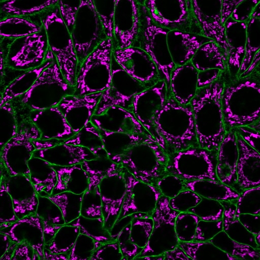 HeLa cells co-stained with CellBrite® Steady 488 (green) and MitoView™ 633 mitochondrial stain (magenta).
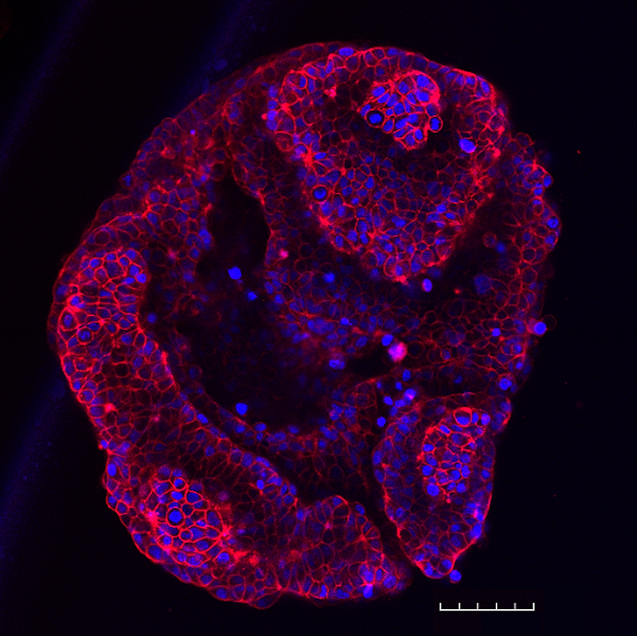 T47-D spheroid stained with NucSpot® Live 650 (nuclei, blue) and CellBrite® Steady 594 (membranes, red). T47-D cells were plated at 5,000 cells per well in a LICORbio 3D Essentials Kit ULA 96-well plate and cultured for 48 hours. The spheroid was stained with dyes in complete medium for 30 minutes without a wash step and imaged on an Evident FV4000 confocal microscope with 20X objective to create a tile-stitched 3D projection image; scale bar: 100 um.
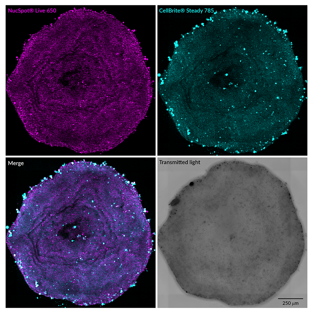 T47-D spheroid stained with NucSpot® Live 650 (nuclei, magenta) and CellBrite® Steady 785 (membranes, cyan). T47-D cells were plated at 20,000 cells per well in a LICORbio 3D Essentials Kit ULA 96-well plate and cultured for 48 hours. The spheroid was stained with dyes in complete medium for 24 hours without a wash step, then imaged on an Evident FV4000 confocal microscope with 20X objective to create a tile-stitched 3D projection image; scale bar: 250 um.
 Enriched EVs were stained with 0.5 uM CellBrite® Steady 405 (right). Specific staining was seen, compared with CellBrite® Steady 405 in buffer (left). EVs were detected on a Cytoflex LX flow cytometer in the Pacific Blue channel.
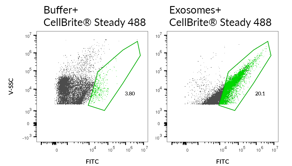 Enriched EVs were stained with 0.5 uM CellBrite Steady® 488 (right). Specific staining was seen, compared with CellBrite® Steady 488 in buffer (left). EVs were detected on a Cytoflex LX flow cytometer in the FITC channel.
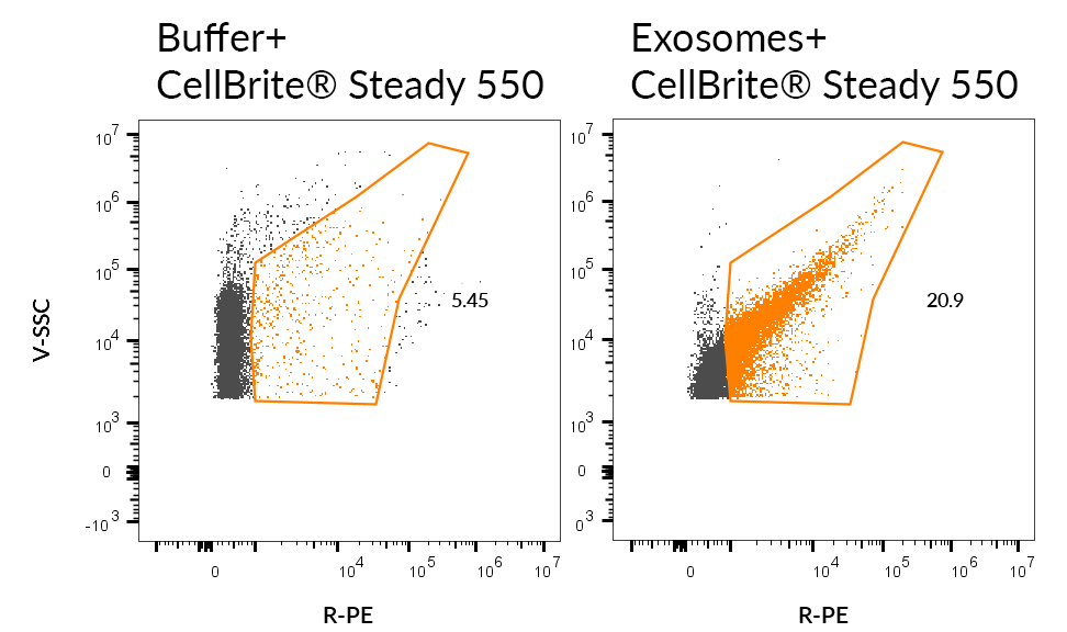 Enriched EVs were stained with 0.5 uM CellBrite® Steady 550 (right). Specific staining was seen, compared with CellBrite® Steady 550 in buffer (left). EVs were detected on a Cytoflex LX flow cytometer in the PE channel.
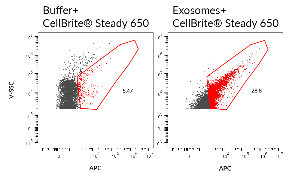 Enriched EVs were stained with 0.5 uM CellBrite® Steady 650 (right). Specific staining was seen, compared with CellBrite® Steady 650 in buffer (left). EVs were detected on a Cytoflex LX flow cytometer in the APC channel.
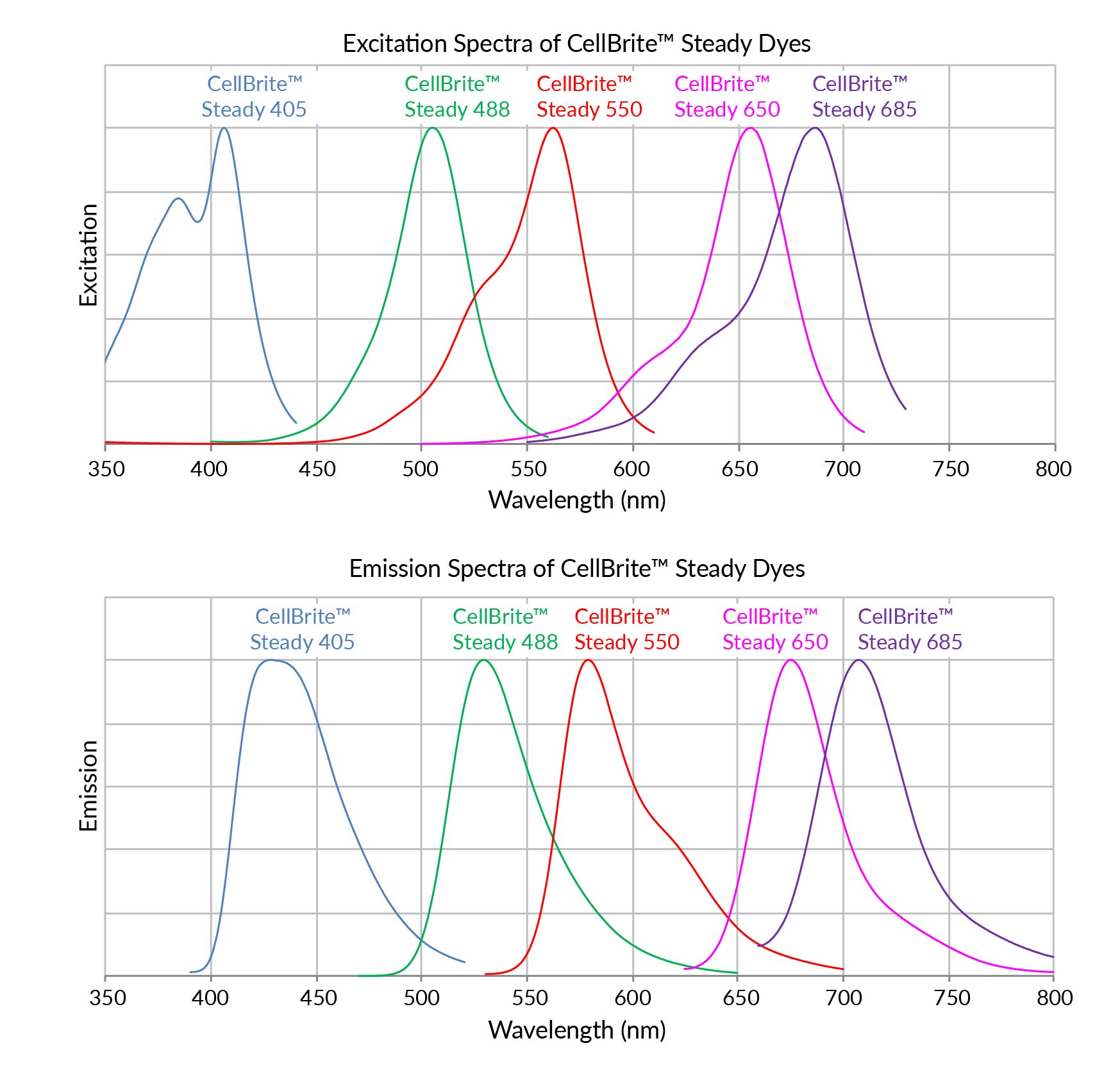 Excitation and emission spectra of CellBrite™ Steady Dyes in methanol.
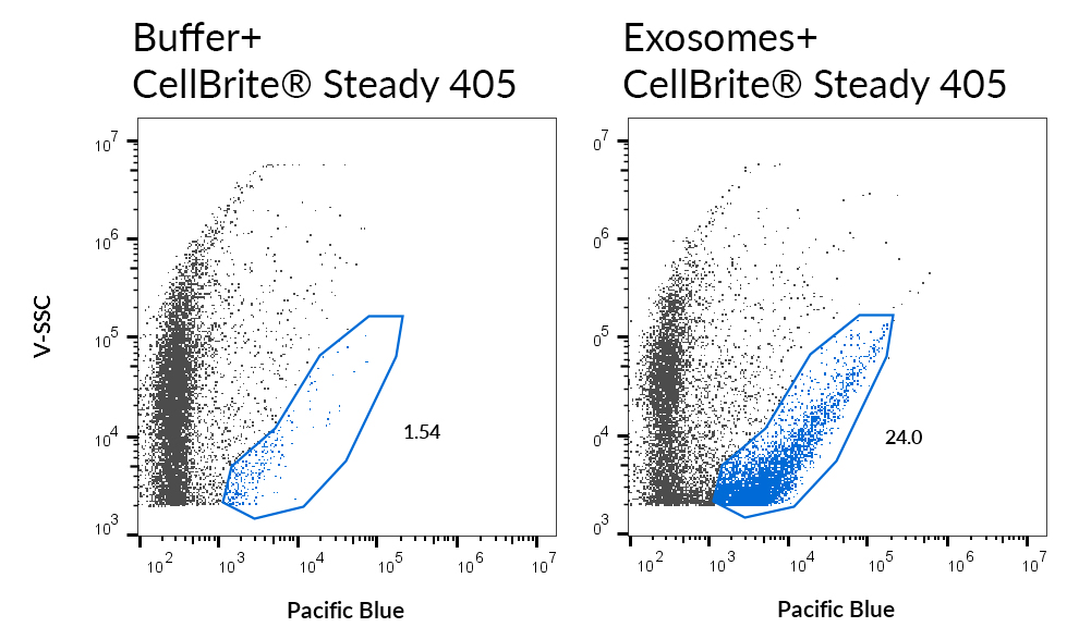 SEC-purified MCF-7-derived exosomes were stained with 0.02X CellBrite® Steady 405 (right). Specific staining was seen, compared with CellBrite® Steady 405 in buffer (left). EVs were detected on a Cytoflex LX flow cytometer in the Pacific Blue channel.
Product Description
CellBrite® Steady Membrane Staining Kits allow fluorescence imaging of cell surface for up to several days in culture. The CellBrite® Steady Dyes are unique fluorescent membrane probes that distribute between the cell surface and intracellular compartments, so cells retain cell surface staining over time. With the use of CellBrite® Steady Enhancer, intracellular staining can be reduced or eliminated for imaging of cell outlines or boundaries.
- Retained on cell surface, unlike other membrane dyes that are rapidly internalized
- Image live cell surface membranes for 24 hours or longer
- Image in your preferred channel with 9 color options from blue to near-IR
- Optional Enhancer masks intracellular signal for selective imaging of cell surface
- Rapid, even staining in complete cell culture medium
- Dye colors from blue to near-IR, with STORM-compatible options
Unlike other membrane/cell surface stains that are rapidly lost from the cell surface by endocytosis after labeling, CellBrite® Steady Dyes equilibrate between intracellular compartments and the plasma membrane. Cells retain surface staining in addition to intracellular staining over the course of hours to days in culture. CellBrite® Steady Enhancer is an optional reagent included in the kits that can be used to mask intracellular fluorescence of CellBrite® Steady Dyes, for more selective visualization of cell boundaries.
Kit Components
Kit sizes are based on 200 uL labeling volume, actual number of labelings may vary based on culture chamber size/staining volume.
- Trial size kit (100 labelings):
- CellBrite® Steady Dye, 1000X in DMSO, 20 uL
- CellBrite® Steady Enhancer, 1000X in DMSO, 20 uL
- Full size kit (500 labelings):
- CellBrite® Steady Dye, 1000X in DMSO, 100 uL
- CellBrite® Steady Enhancer, 1000X in DMSO, 100 uL
Robust & Consistent Long-term Imaging: CellBrite® Steady Outperforms Other Membrane Dyes in Signal Stability
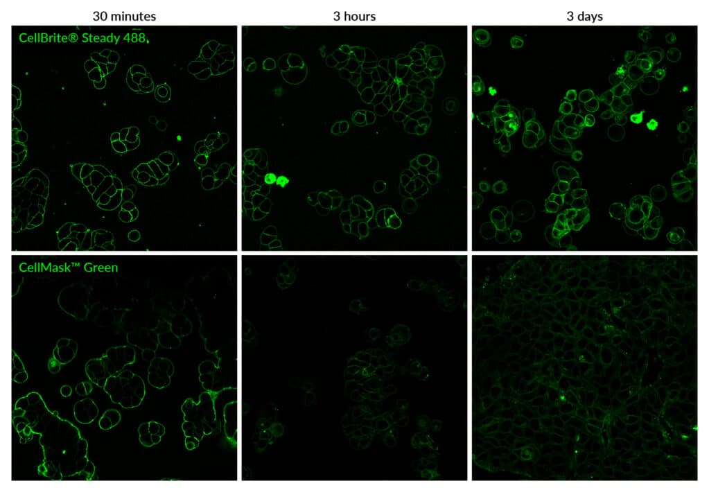
CellBrite® Steady staining is more stable than CellMask™ Green for real-time live cell imaging. Live MCF7 cells were stained with 1X CellBrite™ Steady 488 with CellBrite® Steady Enhancer in complete culture medium at 37°C without a wash step. Cells were cultured for 72 hours after staining and imaged at the time points shown. For CellMask™ Green staining, cell were stained according to the manufacturer's recommendations. Cells were stained with dye diluted 1:1000 in HBSS buffer for 10 minutes at 37°C, then rinsed in buffer, and then fresh culture medium was added. Cells were cultured for 72 hours and imaged at the time points shown.
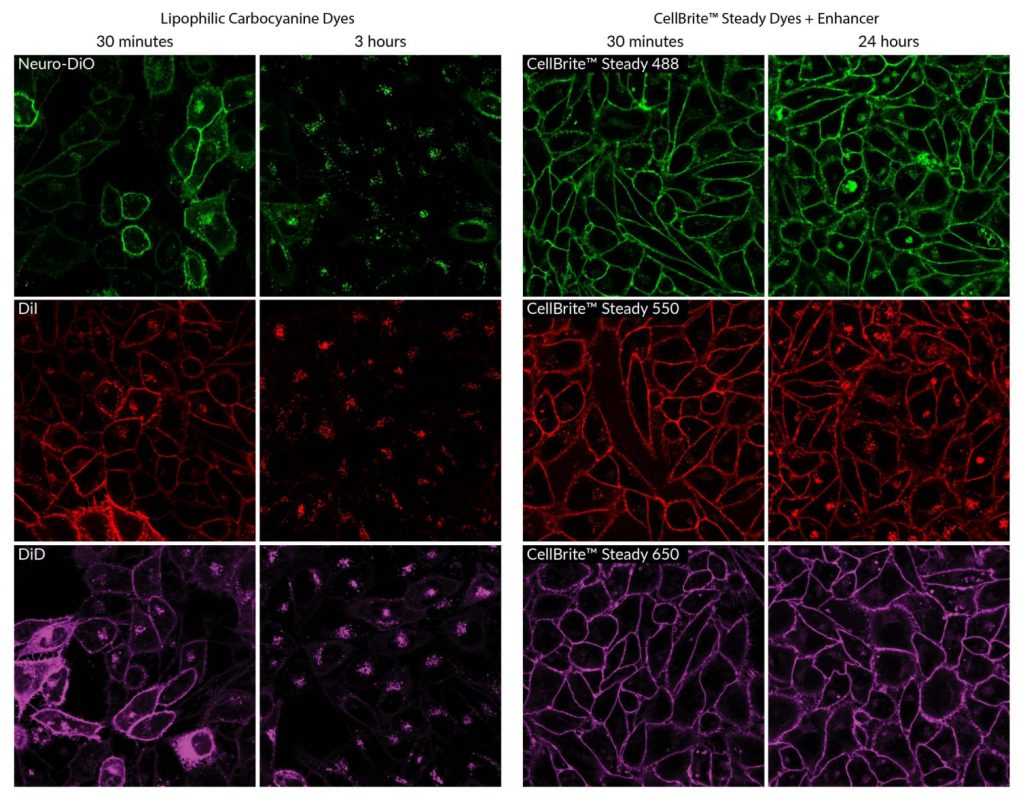
Live HeLa cells stained with the lipophilic carbocyanine dyes Neuro-DiO, DiI, or DiD or CellBrite® Steady Membrane Dyes and Enhancer. Lipophilic carbocyanine dyes are rapidly internalized into intracellular compartments, so surface staining must be imaged within short time frames. CellBrite® Steady Dyes stain cell membranes evenly, and surface staining can be clearly imaged for 24 hours with the use of Enhancer.
Image in Your Preferred Channel with 9 Color Options from Blue to Near-IR
Live MCF7 cells stained with CellBrite® Steady dyes for 30 minutes and imaged by confocal microscopy without a wash step.
Selecting a CellBrite® Steady Dye
CellBrite® Steady Dyes are available in colors from blue to near-infrared. CellBrite® Steady 550, 650, and 685 fluorophores are compatible with super-resolution imaging by STORM. Masking of intracellular fluorescence by CellBrite® Steady Enhancer tends to show more complete masking of intracellular fluorescence for far-red CellBrite® Steady 650 and CellBrite® Steady 685 dyes.
Tips for Success
Washing after staining is optional for imaging by confocal microscopy, but is required for imaging by epifluorescence. For dyes with fluorescence in the visible range (405, 488, and 550), staining may or may not be visible through the microscope eyepieces without washing, so you may need to use brightfield or another marker to focus on cells initially for no-wash confocal imaging.
CellBrite® Steady Dyes have low toxicity and can be continuously incubated in cell culture medium. Enhancer may be toxic to some cell types, especially at higher concentrations. Enhancer may be incubated together with membrane dyes or added after staining to minimize any potential effect of Enhancer on cells. See the Product Protocol for details.
Find the Right Stain for Your Application
CellBrite® Steady Dyes must be used for staining live cells. They cannot be used to stain cells that are already fixed (the dyes primarily label intracellular membranes in fixed cells). Our original CellBrite® Cytoplasmic Membrane Dyes can be used to stain cells after fixation. To find the right stain for your application, see our Membrane & Cell Surface Stains Comparison, or download our Membrane & Surface Stains Brochure.
Membrane staining with CellBrite® Steady Dyes is retained immediately after fixation with formaldehyde, but does not tolerate methanol or detergent. If cells are stored after formaldehyde fixation, over time the dyes will redistribute to stain cytoplasmic structures. Enhancer does not tolerate fixation; if Enhancer-treated cells are fixed after staining, masking will be lost and intracellular fluorescence will return. For fixable cell surface staining that tolerates permeabilization for immunofluorescence staining, we recommend our CellBrite® Fix Membrane Stains or MemBrite® Fix Cell Surface Staining Kits.
CellBrite® Steady Dyes readily transfer between cells, and are not recommended for cell tracking, co-culture, or transplantation studies. Our stable, non-toxic ViaFluor® SE Cell Proliferation Kits can be used to covalently label cells for long-term tracking by microscopy or flow cytometry. See our Tech Tip: Using ViaFluor® SE Stains for Cell Tracing and Co-Culture to learn more.
CellBrite® Steady Dyes have been validated for labeling of purified extracellular vesicles (EVs) and exosomes. However, our tests have shown CellBrite® Steady to bind non-specifically to bead-bound exosomes. For optimal labeling of both purified and bead-bound exosomes we recommend our ExoBrite™ True EV Membrane Stains. Learn more about our ExoBrite™ stains and validated antibodies for Exosome & EV Labeling.
Watch our video where Technical Applications Scientist II, Jacqueline Steenhuis PhD answers your top questions about Biotium's various membrane stains for fluorescence microscopy.
For additional support or product recommendations, contact us at [email protected].
CellBrite® Steady Membrane Staining Kits
| Product | Ex/Em | Laser Line(s) (nm) | Detection Channel | Size | Catalog Number |
|---|---|---|---|---|---|
| CellBrite® Steady 405 Membrane Staining Kit | 406/428 nm | 405 | DAPI | 100 Labelings | 30105-T |
| 500 Labelings | 30105 | ||||
| CellBrite® Steady 488 Membrane Staining Kit | 505/529 nm | 488 | FITC | 100 Labelings | 30106-T |
| 500 Labelings | 30106 | ||||
| CellBrite® Steady 550 Membrane Staining Kit | 562/579 nm | 555 or 561 | TRITC or Rhodamine | 100 Labelings | 30107-T |
| 500 Labelings | 30107 | ||||
| CellBrite® Steady 568 Membrane Staining Kit | 569/588 nm | 555 or 561 | Alexa Fluor® 568 | 100 Labelings | 30150-T |
| 500 Labelings | 30150 | ||||
| CellBrite® Steady 594 Membrane Staining Kit | 580/615 nm | 555, 561, or 594 | Texas Red® | 100 Labelings | 30142-T |
| 500 Labelings | 30142 | ||||
| CellBrite® Steady 650 Membrane Staining Kit | 656/676 nm | 640 | Alexa Fluor® 647 | 100 Labelings | 30108-T |
| 500 Labelings | 30108 | ||||
| CellBrite® Steady 685 Membrane Staining Kit | 686/708 nm | 685 | Alexa Fluor® 680 | 100 Labelings | 30109-T |
| 500 Labelings | 30109 | ||||
| CellBrite® Steady 750 Membrane Staining Kit | 755/789 nm | 730 | Alexa Fluor® 750 | 100 Labelings | 30141-T |
| 500 Labelings | 30141 | ||||
| CellBrite® Steady 785 Membrane Staining Kit | 788/815 m | 785 | Alexa Fluor® 790 | 100 Labelings | 30152-T |
| 500 Labelings | 30152 |
ALEXA FLUOR and TEXAS RED are registered trademarks of Thermo Fisher Scientific.
Technology

Highlighted Citations

Tech Tips


 Powered by Bioz