

VPS35 Antibody, Clone 8A3: ATTO 488

Referencia MBS8005639-0,1mg
embalaje : 0.1mg
Marca : MyBiosource
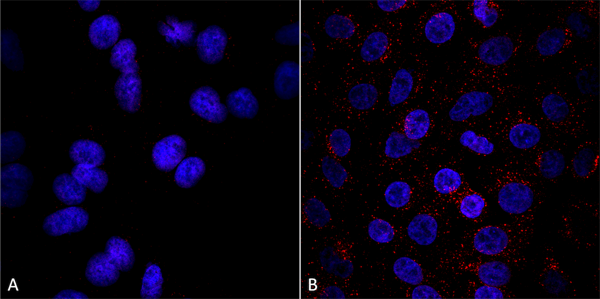
Immunocytochemistry/Immunofluorescence analysis using Mouse Anti-VPS35 Monoclonal Antibody, Clone 8A3. Tissue: A549 cells. Species: Human. Primary Antibody: Mouse Anti-VPS35 Monoclonal Antibody at 1:5 (tissue culture supernatant). Secondary Antibody: Donkey anti-mouse: Alexa Fluor 594 at 1:4000 in 0.2% BSA PBS. Counterstain: DAPI. Localization: Vesicles. A) VPS35 KO A549 cells B) WT A549 cells. Courtesy of: Dario Alessi Lab, University of Dundee.
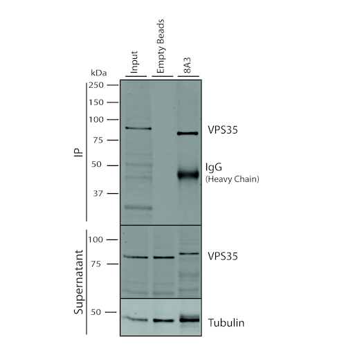
Immunoprecipitation analysis using Mouse Anti-VPS35 Monoclonal Antibody, Clone 8A3. Tissue: A549 cells. Species: Human. Primary Antibody: Mouse Anti-VPS35 Monoclonal Antibody. 500 uL cell culture supernatants were incubated with 10 uL of Protein A/G resin beads for 1 hour at 4 degree C. clone 8A3 depletes VPS35 from the A549 cell extract..
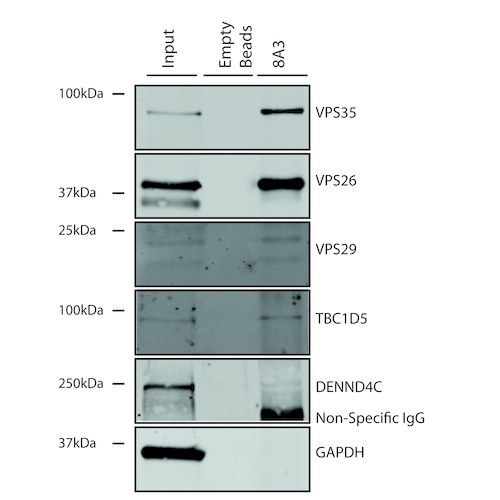
Co-Immunoprecipitation analysis of known interactors of VPS35 using Mouse Anti-VPS35 Monoclonal Antibody, Clone 8A3. Tissue: A549 cells. Species: Human. Primary Antibody: Mouse Anti-VPS35 Monoclonal Antibody.
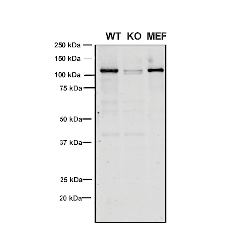
Western Blot analysis of Human, Mouse A549, MEF showing detection of VPS35 protein using Mouse Anti-VPS35 Monoclonal Antibody, Clone 8A3. Lane 1: Molecular Weight Ladder. Lane 2: VPS35 KO A549 cells. Lane 3: mouse embryonic fibroblast cells.. Load: 8 ug each A549 and MEF. Primary Antibody: Mouse Anti-VPS35 Monoclonal Antibody at 1:5 (tissue culture supernatant). Secondary Antibody: Donkey anti-mouse IRDye 800CW at 1:25000 in TBS-T.
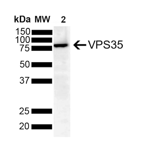
Western Blot analysis of Human SH-SY5Y showing detection of VPS35 protein using Mouse Anti-VPS35 Monoclonal Antibody, Clone 8A3. Lane 1: Molecular Weight Ladder. Lane 2: SH-SY5Y (10 ug). Load: 10 ug. Block: 5% Skim Milk powder in TBST. Primary Antibody: Mouse Anti-VPS35 Monoclonal Antibody at 1:1000 for 2 hours at RT with shaking. Secondary Antibody: Goat anti-mouse IgG:HRP at 1:4000 for 1 hour at RT with shaking. Color Development: Chemiluminescent for HRP (Moss) for 5 min in RT.
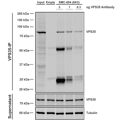
Immunoprecipitation analysis using Mouse Anti-VPS35 Monoclonal Antibody, Clone 8A3. Tissue: A549 cells. Species: Human. Primary Antibody: Mouse Anti-VPS35 Monoclonal Antibody. Three amounts of (3, 1 and 0.3 ug) were non-covalently coupled to 10uL of A/G sepharose beads for 1 hour at 4 degree C and next incubated with 250ug of A549 lysate for 2 hours at 4 degree C.
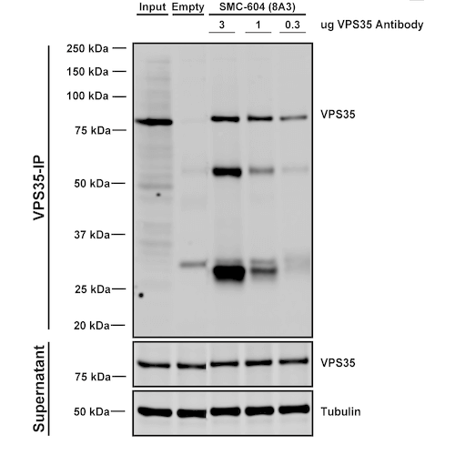
Immunoprecipitation analysis using Mouse Anti-VPS35 Monoclonal Antibody, Clone 8A3. Tissue: embryonic fibroblast. Species: Mouse. Primary Antibody: Mouse Anti-VPS35 Monoclonal Antibody. Three amounts of (3, 1 and 0.3 ug) were non-covalently coupled to 10uL of A/G sepharose beads for 1 hour at 4 degree C and next incubated with 250ug of MEF lysate for 2 hours at 4 degree C.
VPS35 Antibody – Mouse Monoclonal
For Research Use Only. Not for use in diagnostic procedures. Catalog #: MBS8005639Vacuolar Protein Sorter-35 (VPS35) is a component of the retromer complex, which is essential for endosome-to-Golgi retrieval of membrane proteins. VPS35 mutations such as D620N have been linked to Parkinson's Disease (PD) (1,2) and affect retromer function,… More details
Product Overview
Online Datasheet in PDFProduct Name
VPS35, Monoclonal Antibody
Full Product Name
VPS35 Antibody, Clone 8A3: ATTO 488
Product Gene Name
anti-VPS35 antibody
Product Synonym Names
Mouse Anti-Human VPS35 Monoclonal IgG1; VPS35 Antibody: ATTO 488; Vacuolar protein sorting-associated protein 35; MEM3; PARK17; VPS35 retromer complex component; maternal-embryonic 3; vesicle protein sortin 35; TCCCTA00141; FLJ10752
Clonality
Monoclonal
Isotype
IgG1
Clone Number
8A3
Host
Mouse
Species Reactivity
Human, Mouse, Rat
Form/Format
PBS pH 7.4, 50% Glycerol, 0.09% Sodium Azide
Purity/Purification
Protein G Purified
Concentration
1 mg/ml (lot specific)
Specificity
Detects ~92 kDa.
Preparation and Storage
Store at -20 degree C.
Matching Pairs
Unconjugated Antibody: VPS35 Clone #8A3 (MBS8005637)
ATTO 488 Conjugated Antibody: VPS35 Clone #8A3 (MBS8005639)
ATTO 488 Conjugated Antibody: VPS35 Clone #8A3 (MBS8005639)
OMIM
601501
Immunogen Species
Human
Immunogen
Full length recombinant human VSP35
Conjugate
ATTO 488
Cellular Localization
Endosome; Lysosome; Vesicles; Cytoplasm; Membrane
Tissue Specificity
Ubiquitous. Highly expressed in brain, colon, heart, kidney, ovary, placenta, skeletal muscle, small intestine, testis, thymus.
Product Categories/Family for anti-VPS35 antibody
Cell Signaling; Neuroscience |Neurodegeneration; Parkinson's Disease; Membrane Trafficking Proteins; Golgi Proteins; Protein Trafficking; Alzheimer's Disease; Antibodies
Applications Tested/Suitable for anti-VPS35 antibody
Western Blot (WB), Immunocytochemistry (ICC), Immunofluorescence (IF), Immunoprecipitation (IP)
Application Notes for anti-VPS35 antibody
WB: 1:1000
ICC/IF: 1:200
IP: 1:200
ICC/IF: 1:200
IP: 1:200
Immunocytochemistry (ICC) of anti-VPS35 antibody
Immunocytochemistry/Immunofluorescence analysis using Mouse Anti-VPS35 Monoclonal Antibody, Clone 8A3. Tissue: A549 cells. Species: Human. Primary Antibody: Mouse Anti-VPS35 Monoclonal Antibody at 1:5 (tissue culture supernatant). Secondary Antibody: Donkey anti-mouse: Alexa Fluor 594 at 1:4000 in 0.2% BSA PBS. Counterstain: DAPI. Localization: Vesicles. A) VPS35 KO A549 cells B) WT A549 cells. Courtesy of: Dario Alessi Lab, University of Dundee.
Immunoprecipitation (IP) of anti-VPS35 antibody
Immunoprecipitation analysis using Mouse Anti-VPS35 Monoclonal Antibody, Clone 8A3. Tissue: A549 cells. Species: Human. Primary Antibody: Mouse Anti-VPS35 Monoclonal Antibody. 500 uL cell culture supernatants were incubated with 10 uL of Protein A/G resin beads for 1 hour at 4 degree C. clone 8A3 depletes VPS35 from the A549 cell extract..
Western Blot (WB) of anti-VPS35 antibody
Western Blot analysis of Human, Mouse A549, MEF showing detection of VPS35 protein using Mouse Anti-VPS35 Monoclonal Antibody, Clone 8A3. Lane 1: Molecular Weight Ladder. Lane 2: VPS35 KO A549 cells. Lane 3: mouse embryonic fibroblast cells.. Load: 8 ug each A549 and MEF. Primary Antibody: Mouse Anti-VPS35 Monoclonal Antibody at 1:5 (tissue culture supernatant). Secondary Antibody: Donkey anti-mouse IRDye 800CW at 1:25000 in TBS-T.
Western Blot (WB) of anti-VPS35 antibody
Western Blot analysis of Human SH-SY5Y showing detection of VPS35 protein using Mouse Anti-VPS35 Monoclonal Antibody, Clone 8A3. Lane 1: Molecular Weight Ladder. Lane 2: SH-SY5Y (10 ug). Load: 10 ug. Block: 5% Skim Milk powder in TBST. Primary Antibody: Mouse Anti-VPS35 Monoclonal Antibody at 1:1000 for 2 hours at RT with shaking. Secondary Antibody: Goat anti-mouse IgG:HRP at 1:4000 for 1 hour at RT with shaking. Color Development: Chemiluminescent for HRP (Moss) for 5 min in RT.
Immunoprecipitation (IP) of anti-VPS35 antibody
Immunoprecipitation analysis using Mouse Anti-VPS35 Monoclonal Antibody, Clone 8A3. Tissue: A549 cells. Species: Human. Primary Antibody: Mouse Anti-VPS35 Monoclonal Antibody. Three amounts of (3, 1 and 0.3 ug) were non-covalently coupled to 10uL of A/G sepharose beads for 1 hour at 4 degree C and next incubated with 250ug of A549 lysate for 2 hours at 4 degree C.
Immunoprecipitation (IP) of anti-VPS35 antibody
Immunoprecipitation analysis using Mouse Anti-VPS35 Monoclonal Antibody, Clone 8A3. Tissue: embryonic fibroblast. Species: Mouse. Primary Antibody: Mouse Anti-VPS35 Monoclonal Antibody. Three amounts of (3, 1 and 0.3 ug) were non-covalently coupled to 10uL of A/G sepharose beads for 1 hour at 4 degree C and next incubated with 250ug of MEF lysate for 2 hours at 4 degree C.
Product References and Citations for anti-VPS35 antibody
1. Vilarino-Guell, C. et al. (2011) Am J Hum Genet 89:162-167. 2. Zimprich, A. et al. (2011) Am J Hum Genet 89:168-175. 3. Rahman, A.A., Morrison, B.E. (2019) Neurosci 401:1-10.
Other Notes
Small volumes of anti-VPS35 antibody vial(s) may occasionally become entrapped in the seal of the product vial during shipment and storage. If necessary, briefly centrifuge the vial on a tabletop centrifuge to dislodge any liquid in the container`s cap. Certain products may require to ship with dry ice and additional dry ice fee may apply.
Searchable Terms for VPS35 purchase
MBS8005639 is an antibody targeting VPS35. Also known as VPS35 Antibody, Clone 8A3: ATTO 488. Reactive in Human, Mouse, Rat. Host: Mouse.
Additional Details
NCBI/Uniprot data below describe general gene information for VPS35. It may not necessarily be applicable to this product.
NCBI Official Full Name
vacuolar protein sorting-associated protein 35
NCBI Official Synonym Full Names
VPS35 retromer complex component
NCBI Official Symbol
VPS35
NCBI Official Synonym Symbols
MEM3; PARK17
UniProt Protein Name
Vacuolar protein sorting-associated protein 35
UniProt Synonym Protein Names
Maternal-embryonic 3; Vesicle protein sorting 35
Protein Family
Vacuolar protein sorting-associated protein
UniProt Gene Name
VPS35
UniProt Synonym Gene Names
MEM3; hVPS35
UniProt Entry Name
VPS35_HUMAN
NCBI Protein Information
vacuolar protein sorting-associated protein 35
NCBI Summary for VPS35
This gene belongs to a group of vacuolar protein sorting (VPS) genes. The encoded protein is a component of a large multimeric complex, termed the retromer complex, involved in retrograde transport of proteins from endosomes to the trans-Golgi network. The close structural similarity between the yeast and human proteins that make up this complex suggests a similarity in function. Expression studies in yeast and mammalian cells indicate that this protein interacts directly with VPS35, which serves as the core of the retromer complex. [provided by RefSeq, Jul 2008]
Show more Precautions
All of MyBioSource's Products are for scientific laboratory research purposes and are not for diagnostic, therapeutics, prophylactic or in vivo use. Through your purchase, you expressly represent and warrant to MyBioSource that you will properly test and use any Products purchased from MyBioSource in accordance with industry standards. MyBioSource and its authorized distributors reserve the right to refuse to process any order where we reasonably believe that the intended use will fall outside of our acceptable guidelines.
Disclaimer
While every efforts were made to ensure the accuracy of the information provided in this datasheet, MyBioSource will not be liable for any omissions or errors contained herein. MyBioSource reserves the right to make changes to this datasheet at any time without prior notice.
It is the responsibility of the customer to report product performance issues to MyBioSource within 30 days of receipt of the product. Please visit our Terms & Conditions page for more information.
It is the responsibility of the customer to report product performance issues to MyBioSource within 30 days of receipt of the product. Please visit our Terms & Conditions page for more information.
Product Note
Select online data sheet information is drawn from bioinformatics databases, occasionally resulting in ambiguous or non-relevant product information. It is the responsibility of the customer to review, verify, and evaluate the information to make sure it matches their requirements before purchasing the kit. Our ELISA Kit assays are dynamic research tools and sometimes they may be updated and improved. If the format of this assay is important to you then please request the current manual or contact our technical support team with a presales inquiry before placing an order. We will confirm the current details of the assay. We cannot guarantee the sample manual posted online is the most current manual, it is intended to serve as an example only. Please refer to the instructions For Use provided with the assay kit for precise details.
Product PubMed Publications
Products associated with anti-VPS35 antibody
| Reference Product | PubMed Publications |
|---|---|
| VPS29 antibody | >31 publications with VPS35 and VPS29 |
| SNX1 antibody | >15 publications with VPS35 and SNX1 |
| VPS26B antibody | >5 publications with VPS35 and VPS26B |
Pathways associated with anti-VPS35 antibody
| Products by Pathway | Pathway Diagram |
|---|---|
| Endocytosis Pathway antibodies | Endocytosis Pathway Diagram |
| Endocytosis Pathway antibodies | Endocytosis Pathway Diagram |
| Signal Transduction Pathway antibodies | Signal Transduction Pathway Diagram |
| Signaling By Wnt Pathway antibodies | Signaling By Wnt Pathway Diagram |
| WNT Ligand Biogenesis And Trafficking Pathway antibodies | WNT Ligand Biogenesis And Trafficking Pathway Diagram |
Diseases associated with anti-VPS35 antibody
| Disease Name | Pubmed Publications |
|---|---|
| Colorectal Neoplasms Antibodies | >1 publications with VPS35 and Colorectal Neoplasms |
| Lung Neoplasms Antibodies | >1 publications with VPS35 and Lung Neoplasms |
| Adenocarcinoma Antibodies | >1 publications with VPS35 and Adenocarcinoma |
| Carcinoma, Squamous Cell Antibodies | >1 publications with VPS35 and Carcinoma, Squamous Cell |
| Carcinoma Antibodies | >1 publications with VPS35 and Carcinoma |
| Neoplasms Antibodies | >1 publications with VPS35 and Neoplasms |
Organs/Tissues associated with anti-VPS35 antibody
| Organ/Tissue Name | Pubmed Publications |
|---|---|
| Kidney Antibodies | >4 publications with VPS35 and Kidney |
| Brain Antibodies | >4 publications with VPS35 and Brain |
| Heart Antibodies | >3 publications with VPS35 and Heart |
| Intestine Antibodies | >2 publications with VPS35 and Intestine |
| Testis Antibodies | >2 publications with VPS35 and Testis |
| Blood Antibodies | >2 publications with VPS35 and Blood |
| Spleen Antibodies | >1 publications with VPS35 and Spleen |
| Liver Antibodies | >1 publications with VPS35 and Liver |
| Embryonic Tissue Antibodies | >1 publications with VPS35 and Embryonic Tissue |
| Placenta Antibodies | >1 publications with VPS35 and Placenta |