MDM2 antibody

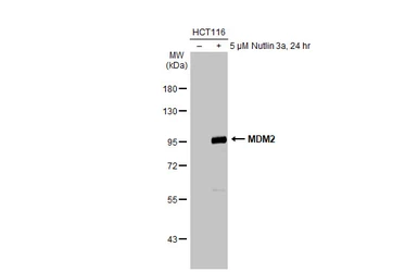
Untreated (–) and treated (+) HCT-116 whole cell extracts (30 μg) were separated by 7.5% SDS-PAGE, and the membrane was blotted with MDM2 antibody (GTX100531) diluted at 1:1000. The HRP-conjugated anti-rabbit IgG antibody (GTX213110-01) was used to detect the primary antibody.
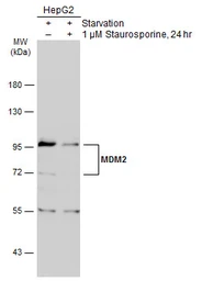
Untreated (–) and treated (+) HepG2 whole cell extracts (30 μg) were separated by 7.5% SDS-PAGE, and the membrane was blotted with MDM2 antibody (GTX100531) diluted at 1:3000. The HRP-conjugated anti-rabbit IgG antibody (GTX213110-01) was used to detect the primary antibody.
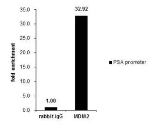
ChIP experiment and primer designs are based on Nucleic Acids Res. 2005 Jan 7;33(1):13-26.
Cross-linked ChIP was performed with PC-3 chromatin extract and 5 μg of either control rabbit IgG or anti-MDM2 antibody. The precipitated DNA was detected by PCR with primer set targeting to PSA promoter.
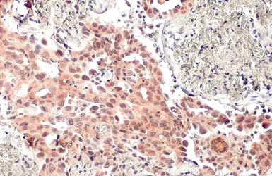
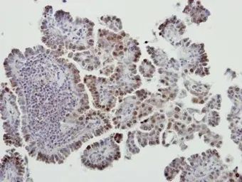
MDM2 antibody detects MDM2 protein at nucleus by immunohistochemical analysis.Sample: Paraffin-embedded human lung cancer.MDM2 stained by MDM2 antibody (GTX100531) diluted at 1:500.Antigen Retrieval: Citrate buffer, pH 6.0, 15 min
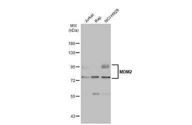
Various whole cell extracts (30 μg) were separated by 7.5% SDS-PAGE, and the membrane was blotted with MDM2 antibody (GTX100531) diluted at 1:1000. The HRP-conjugated anti-rabbit IgG antibody (GTX213110-01) was used to detect the primary antibody.
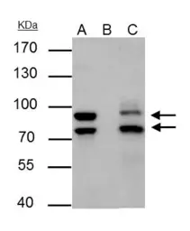
MDM2 antibody immunoprecipitates MDM2 protein in IP experiments.
IP samples: Jurkat whole cell extract
A. 30 μg Jurkat cell whole cell extract
B. Control with 4 μg of preimmune Rabbit IgG
C. Immunoprecipitation of MDM2 protein by 4 μg MDM2 antibody (GTX100531)
7.5 % SDS-PAGE
The immunoprecipitated MDM2 protein was detected by MDM2 antibody (GTX100531) diluted at 1:500.
[EasyBlot anti-rabbit IgG (GTX221666-01) was used as a secondary reagent]
Immunohistochemical analysis of paraffin-embedded human lung cancer, using MDM2(GTX100531) antibody at 1:500 dilution.
Antigen Retrieval: Trilogy™ (EDTA based, pH 8.0) buffer, 15min
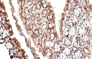
MDM2 antibody detects MDM2 protein at nucleus by immunohistochemical analysis.Sample: Paraffin-embedded mouse colon.MDM2 stained by MDM2 antibody (GTX100531) diluted at 1:500.Antigen Retrieval: Citrate buffer, pH 6.0, 15 min
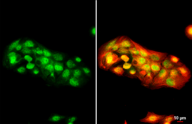
MDM2 antibody detects MDM2 protein at cytoplasm and nucleus by immunofluorescent analysis.
Sample: HepG2 cells were fixed in 4% paraformaldehyde at RT for 15 min.
Green: MDM2 stained by MDM2 antibody (GTX100531) diluted at 1:500.
Red: alpha Tubulin, a cytoskeleton marker, stained by alpha Tubulin antibody [GT114] (GTX628802) diluted at 1:1000.
Scale bar= 10μm.